 Movie
Movie Controller


+ Open data

- Basic information
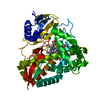
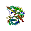
| Entry | Database: PDB / ID: 2pvi | ||||||
|---|---|---|---|---|---|---|---|
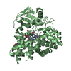
| Title | PVUII ENDONUCLEASE COMPLEXED TO AN IODINATED COGNATE DNA | ||||||
 Components Components |
| ||||||
 Keywords Keywords | HYDROLASE/DNA / COMPLEX (RESTRICTION ENDONUCLEASE-DNA) / HYDROLASE-DNA COMPLEX | ||||||
| Function / homology |  Function and homology information Function and homology informationtype II site-specific deoxyribonuclease / type II site-specific deoxyribonuclease activity / DNA restriction-modification system / DNA binding / metal ion binding Similarity search - Function | ||||||
| Biological species |  Proteus vulgaris (bacteria) Proteus vulgaris (bacteria) | ||||||
| Method |  X-RAY DIFFRACTION / SYNCHROTRON / MOLECULAR REPLACEMENT / Resolution: 1.76 Å X-RAY DIFFRACTION / SYNCHROTRON / MOLECULAR REPLACEMENT / Resolution: 1.76 Å | ||||||
 Authors Authors | Horton, J. / Cheng, X. | ||||||
 Citation Citation | Journal: J.Biol.Chem. / Year: 1998 Title: How is modification of the DNA substrate recognized by the PvuII restriction endonuclease? Authors: Horton, J.R. / Bonventre, J. / Cheng, X. #1: Journal: Proteins / Year: 1994Title: Expression, Purification, and Crystallization of Restriction Endonuclease PvuII with DNA Containing its Recognition Site Authors: Balendiran, K. / Bonventre, J. / Knott, R. / Jack, W. / Benner, J. / Schildkraut, I. / Anderson, J.E. #2: Journal: Embo J. / Year: 1994Title: Structure of PvuII Endonuclease with Cognate DNA Authors: Cheng, X. / Balendiran, K. / Schildkraut, I. / Anderson, J.E. | ||||||
| History |
|
- Structure visualization
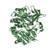
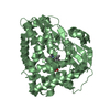
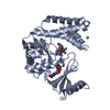
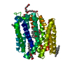
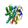
| Structure viewer | Molecule: MolmilJmol/JSmol |
|---|
- Downloads & links
-Download
| PDBx/mmCIF format | 2pvi.cif.gz | 97.1 KB | Display | PDBx/mmCIF format |
|---|---|---|---|---|
| PDB format | pdb2pvi.ent.gz | 70.9 KB | Display | PDB format |
| PDBx/mmJSON format | 2pvi.json.gz | Tree view | PDBx/mmJSON format | |
| Others |  Other downloads Other downloads |
-Validation report
| Arichive directory | https://data.pdbj.org/pub/pdb/validation_reports/pv/2pviftp://data.pdbj.org/pub/pdb/validation_reports/pv/2pvi | HTTPS FTP |
|---|
-Related structure data
| Related structure data |  1pviS S: Starting model for refinement |
|---|---|
| Similar structure data |

-Links
 PDBj
PDBj


- Assembly
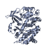
| Deposited unit | 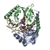
| ||||||||||
|---|---|---|---|---|---|---|---|---|---|---|---|
| 1 |
| ||||||||||
| Unit cell |
|
-Components
| #1: DNA chain | Mass: 4093.481 Da / Num. of mol.: 2 / Source method: obtained synthetically #2: Protein | Mass: 18220.859 Da / Num. of mol.: 2 Source method: isolated from a genetically manipulated source Source: (gene. exp.) Proteus vulgaris (bacteria) / Plasmid: PPR594 / Production host: References: UniProt: P23657, type II site-specific deoxyribonuclease #3: Water | ChemComp-HOH / |  Mass: 18.015 Da / Num. of mol.: 166 / Source method: isolated from a natural source / Formula: H2O Mass: 18.015 Da / Num. of mol.: 166 / Source method: isolated from a natural source / Formula: H2O |
|---|
-Experimental details
-Experiment
| Experiment | Method: X-RAY DIFFRACTION / Number of used crystals: 1 |
|---|
- Sample preparation
| Crystal | Density Matthews: 2.71 Å3/Da / Density % sol: 54 % |
|---|---|
| Crystal grow | Method: vapor diffusion, hanging drop / pH: 4.5 / Details: pH 4.5, VAPOR DIFFUSION, HANGING DROP |
-Data collection
| Diffraction | Mean temperature: 289 K |
|---|---|
| Diffraction source | Source: SYNCHROTRON / Site: NSLS  / Beamline: X12C / Beamline: X12C |
| Detector | Type: MARRESEARCH / Detector: IMAGE PLATE / Details: COLLIMATOR |
| Radiation | Monochromator: SI (111) / Protocol: SINGLE WAVELENGTH / Monochromatic (M) / Laue (L): M / Scattering type: x-ray |
| Radiation wavelength | Relative weight: 1 |
| Reflection | Resolution: 1.76→19.5 Å / % possible obs: 77.3 % / Observed criterion σ(I): 0 / Redundancy: 3.8 % / Rmerge(I) obs: 0.054 / Net I/σ(I): 14.1 |
| Reflection shell | Resolution: 1.76→1.84 Å / % possible all: 23.7 |
- Processing
| Software |
| ||||||||||||||||||||||||||||||||||||||||||||||||||||||||||||
|---|---|---|---|---|---|---|---|---|---|---|---|---|---|---|---|---|---|---|---|---|---|---|---|---|---|---|---|---|---|---|---|---|---|---|---|---|---|---|---|---|---|---|---|---|---|---|---|---|---|---|---|---|---|---|---|---|---|---|---|---|---|
| Refinement | Method to determine structure: MOLECULAR REPLACEMENT Starting model: 1PVI Resolution: 1.76→19.5 Å / Data cutoff high absF: 100000 / Data cutoff low absF: 0.1 / Cross valid method: THROUGHOUT / σ(F): 0 / Details: TWO CONFORMATIONS EXIST FOR HIS A84 AND HIS B84.
| ||||||||||||||||||||||||||||||||||||||||||||||||||||||||||||
| Refinement step | Cycle: LAST / Resolution: 1.76→19.5 Å
| ||||||||||||||||||||||||||||||||||||||||||||||||||||||||||||
| Refine LS restraints |
| ||||||||||||||||||||||||||||||||||||||||||||||||||||||||||||
| LS refinement shell | Resolution: 1.76→1.84 Å / Total num. of bins used: 8 /
| ||||||||||||||||||||||||||||||||||||||||||||||||||||||||||||
| Xplor file |
|
