 Movie
Movie Controller

[English] 日本語
 Yorodumi
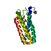
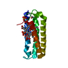
- PDB-2jvw: Solution NMR structure of uncharacterized protein Q5E7H1 from Vib... -
+ Open data

- Basic information
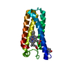
| Entry | Database: PDB / ID: 2jvw | ||||||
|---|---|---|---|---|---|---|---|
| Title | Solution NMR structure of uncharacterized protein Q5E7H1 from Vibrio fischeri. Northeast Structural Genomics target VfR117 | ||||||
 Components Components | Uncharacterized protein | ||||||
 Keywords Keywords |  STRUCTURAL GENOMICS / UNKNOWN FUNCTION / solution NMR structure / alpha helical protein / PSI-2 / Protein Structure Initiative / Northeast Structural Genomics Consortium / NESG STRUCTURAL GENOMICS / UNKNOWN FUNCTION / solution NMR structure / alpha helical protein / PSI-2 / Protein Structure Initiative / Northeast Structural Genomics Consortium / NESG | ||||||
| Function / homology | DNA-binding protein VF530-like / DNA-binding protein VF530 / SAP domain / Transcription Termination Factor Rho, Rna-binding Domain; Chain A, Domain 1 / SAP domain superfamily / double-stranded DNA binding / Orthogonal Bundle / Mainly Alpha / DNA-binding protein VF_0530 Function and homology information Function and homology information | ||||||
| Biological species |  Vibrio fischeri (bacteria) Vibrio fischeri (bacteria) | ||||||
| Method | SOLUTION NMR / simulated annealing | ||||||
 Authors Authors | Aramini, J.M. / Rossi, P. / Wang, D. / Nwosu, C. / Owens, L.A. / Xiao, R. / Liu, J. / Baran, M.C. / Swapna, G.V.T. / Acton, T.B. ...Aramini, J.M. / Rossi, P. / Wang, D. / Nwosu, C. / Owens, L.A. / Xiao, R. / Liu, J. / Baran, M.C. / Swapna, G.V.T. / Acton, T.B. / Rost, B. / Montelione, G.T. / Northeast Structural Genomics Consortium (NESG) | ||||||
 Citation Citation | Journal: Proteins / Year: 2011 Title: Solution NMR structure of VF0530 from Vibrio fischeri reveals a nucleic acid-binding function. Authors: Aramini, J.M. / Rossi, P. / Fischer, M. / Xiao, R. / Acton, T.B. / Montelione, G.T. | ||||||
| History |
|
- Structure visualization
| Structure viewer | Molecule: MolmilJmol/JSmol |
|---|
- Downloads & links
-Download
| PDBx/mmCIF format | 2jvw.cif.gz | 601 KB | Display | PDBx/mmCIF format |
|---|---|---|---|---|
| PDB format | pdb2jvw.ent.gz | 512.2 KB | Display | PDB format |
| PDBx/mmJSON format | 2jvw.json.gz | Tree view | PDBx/mmJSON format | |
| Others |  Other downloads Other downloads |
-Validation report
| Arichive directory | https://data.pdbj.org/pub/pdb/validation_reports/jv/2jvwftp://data.pdbj.org/pub/pdb/validation_reports/jv/2jvw | HTTPS FTP |
|---|
-Related structure data
| Similar structure data | |
|---|---|
| Other databases |

-Links
 PDBj
PDBj
- Assembly
| Deposited unit | 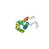
| |||||||||
|---|---|---|---|---|---|---|---|---|---|---|
| 1 |
| |||||||||
| NMR ensembles |
|
-Components
| #1: Protein | Mass: 10746.398 Da / Num. of mol.: 1 Source method: isolated from a genetically manipulated source Source: (gene. exp.) Vibrio fischeri (bacteria) / Species: Aliivibrio fischeri / Strain: ES114 / Gene: VF0530 / Plasmid: VfR117-21.1 / Production host: Escherichia coli (E. coli) / Strain (production host): BL21(DE3)MGK / References: UniProt: Q5E7H1 |
|---|
-Experimental details
-Experiment
| Experiment | Method: SOLUTION NMR | ||||||||||||||||||||||||||||||||||||||||||||||||||||||||||||
|---|---|---|---|---|---|---|---|---|---|---|---|---|---|---|---|---|---|---|---|---|---|---|---|---|---|---|---|---|---|---|---|---|---|---|---|---|---|---|---|---|---|---|---|---|---|---|---|---|---|---|---|---|---|---|---|---|---|---|---|---|---|
| NMR experiment |
| ||||||||||||||||||||||||||||||||||||||||||||||||||||||||||||
| NMR details | Text: THE PROTEIN IS MONOMERIC BY GEL FILTRATION CHROMATOGRAPHY AND STATIC LIGHT SCATTERING. THE STRUCTURE WAS DETERMINED USING TRIPLE RESONANCE NMR SPECTROSCOPY. AUTOMATED BACKBONE ASSIGNMENTS WERE ...Text: THE PROTEIN IS MONOMERIC BY GEL FILTRATION CHROMATOGRAPHY AND STATIC LIGHT SCATTERING. THE STRUCTURE WAS DETERMINED USING TRIPLE RESONANCE NMR SPECTROSCOPY. AUTOMATED BACKBONE ASSIGNMENTS WERE MADE USING AUTOASSIGN, AND THE SIDE CHAIN ASSIGNMENTS WERE COMPLETED MANUALLY. AUTOMATIC NOESY ASSIGNMENTS WERE DETERMINED USING CYANA 2.1. DIHEDRAL ANGLE CONSTRAINTS WERE OBTAINED FROM TALOS. HYDROGEN BOND CONSTRAINTS WERE DETERMINED USING BOTH AUTOSTRUCTURE AND CYANA, AND WERE APPLIED ONLY IN THE FINAL REFINEMENT STAGE (CNS) OF THE STRUCTURE DETERMINATION. COMPLETENESS OF NMR ASSIGNMENTS (EXCLUDING C-TERMINAL HHHHHH): BACKBONE, 98.8%, SIDE CHAIN, 95.3%, AROMATICS, 100%, STEREOSPECIFIC METHYL, 100%, STEREOSPECIFIC SIDE CHAIN NH2: 100%. FINAL STRUCTURE QUALITY FACTORS (FOR RESIDUES 1 TO 82, PSVS 1.3), WHERE ORDERED RESIDUES [S(PHI) + S(PSI) > 1.8] COMPRISE: 15-37,42-43,46-75: (A) RMSD (ORDERED RESIDUES): BB, 0.5, HEAVY ATOM, 1.1. (B) RAMACHANDRAN STATISTICS FOR ORDERED RESIDUES: MOST FAVORED, 93.9, ADDITIONALLY ALLOWED, 6.1%, GENEROUSLY ALLOWED, 0.0%, DISALLOWED, 0.0%. (C) PROCHECK SCORES FOR ORDERED RESIDUES (RAW/Z-): PHI-PSI, 0.17/0.98, ALL, 0.19/1.12. (D) MOLPROBITY CLASH SCORE (RAW/Z-): 14.65/-0.99. (E) RPF SCORES FOR GOODNESS OF FIT TO NOESY DATA (RESIDUES 1-82): RECALL, 0.989, PRECISION, 0.945, F-MEASURE, 0.967, DP-SCORE, 0.810. (F) NUMBER OF CLOSE CONTACTS PER 20 MODELS: 2. THE C-TERMINAL HIS TAG RESIDUES OF THE PROTEIN (HHHHHH) WERE NOT INCLUDED IN THE STRUCTURE CALCULATIONS AND HAVE BEEN OMITTED FROM THIS DEPOSITION. COORDINATES FOR THE FOLLOWING RESIDUES ARE NOT WELL DETERMINED [S(PHI) + S(PSI) < 1.8]: 1-14,38-41,44-45,76-82. |
- Sample preparation
| Details |
| ||||||||||||||||||||||||||||||||||||||||||||||||||||
|---|---|---|---|---|---|---|---|---|---|---|---|---|---|---|---|---|---|---|---|---|---|---|---|---|---|---|---|---|---|---|---|---|---|---|---|---|---|---|---|---|---|---|---|---|---|---|---|---|---|---|---|---|---|
| Sample |
| ||||||||||||||||||||||||||||||||||||||||||||||||||||
| Sample conditions | Ionic strength: 0.1 / pH: 4.5 / Pressure: ambient / Temperature: 293 K |
-NMR measurement
| NMR spectrometer |
|
|---|
- Processing
| NMR software |
| ||||||||||||||||||||||||||||||||||||||||||||
|---|---|---|---|---|---|---|---|---|---|---|---|---|---|---|---|---|---|---|---|---|---|---|---|---|---|---|---|---|---|---|---|---|---|---|---|---|---|---|---|---|---|---|---|---|---|
| Refinement | Method: simulated annealing / Software ordinal: 1 Details: THE FINAL STRUCTURES ARE BASED ON A TOTAL OF 1424 CONFORMATIONALLY-RESTRICTING NOE-DERIVED DISTANCE CONSTRAINTS, 88 DIHEDRAL ANGLE CONSTRAINTS, AND 46 HYDROGEN BOND CONSTRAINTS (19.2 ...Details: THE FINAL STRUCTURES ARE BASED ON A TOTAL OF 1424 CONFORMATIONALLY-RESTRICTING NOE-DERIVED DISTANCE CONSTRAINTS, 88 DIHEDRAL ANGLE CONSTRAINTS, AND 46 HYDROGEN BOND CONSTRAINTS (19.2 CONSTRAINTS PER RESIDUE, 3.3 LONG RANGE CONSTRAINTS PER RESIDUE, COMPUTED FOR RESIDUES 1 TO 82 BY PSVS 1.3). STRUCTURE DETERMINATION WAS PERFORMED ITERATIVELY USING CYANA 2.1. THE 20 STRUCTURES OUT OF 100 WITH THE LOWEST TARGET FUNCTION WERE FURTHER REFINED BY RESTRAINED MOLECULAR DYNAMICS/ENERGY MINIMIZATION IN EXPLICIT WATER (CNS) WITH PARAM19. | ||||||||||||||||||||||||||||||||||||||||||||
| NMR constraints | NOE constraints total: 1424 / NOE intraresidue total count: 489 / NOE long range total count: 270 / NOE medium range total count: 285 / NOE sequential total count: 380 | ||||||||||||||||||||||||||||||||||||||||||||
| NMR representative | Selection criteria: lowest energy | ||||||||||||||||||||||||||||||||||||||||||||
| NMR ensemble | Conformer selection criteria: structures with the lowest energy Conformers calculated total number: 100 / Conformers submitted total number: 20 / Maximum lower distance constraint violation: 0 Å / Maximum torsion angle constraint violation: 1.2 ° / Maximum upper distance constraint violation: 0.32 Å / Torsion angle constraint violation method: PDBStat | ||||||||||||||||||||||||||||||||||||||||||||
| NMR ensemble rms | Distance rms dev: 0.01 Å |
