 Movie
Movie Controller

[English] 日本語
 Yorodumi
- PDB-2ia5: T4 polynucleotide kinase/phosphatase with bound sulfate and magnesium. -
+ Open data

- Basic information
| Entry | Database: PDB / ID: 2ia5 | ||||||
|---|---|---|---|---|---|---|---|
| Title | T4 polynucleotide kinase/phosphatase with bound sulfate and magnesium. | ||||||
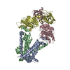
 Components Components | Polynucleotide kinase | ||||||
 Keywords Keywords | TRANSFERASE / Polynucleotide Kinase Phosphatase Sulfate-Complex | ||||||
| Function / homology |  Function and homology information Function and homology informationdeoxynucleotide 3'-phosphatase / deoxynucleotide 3'-phosphatase activity / polynucleotide 5'-hydroxyl-kinase / ATP-dependent polydeoxyribonucleotide 5'-hydroxyl-kinase activity / DNA repair / ATP binding Similarity search - Function | ||||||
| Biological species |  Enterobacteria phage T4 (virus) Enterobacteria phage T4 (virus) | ||||||
| Method |  X-RAY DIFFRACTION / SYNCHROTRON / MOLECULAR REPLACEMENT / Resolution: 2.9 Å X-RAY DIFFRACTION / SYNCHROTRON / MOLECULAR REPLACEMENT / Resolution: 2.9 Å | ||||||
 Authors Authors | Zhu, H. / Smith, P.C. / Wang, L.K. / Lima, C.D. / Shuman, S. | ||||||
 Citation Citation | Journal: Virology / Year: 2007 Title: Structure-function analysis of the 3' phosphatase component of T4 polynucleotide kinase/phosphatase. Authors: Zhu, H. / Smith, P. / Wang, L.K. / Shuman, S. | ||||||
| History |
|
- Structure visualization
| Structure viewer | Molecule: MolmilJmol/JSmol |
|---|
- Downloads & links
-Download
| PDBx/mmCIF format | 2ia5.cif.gz | 721.1 KB | Display | PDBx/mmCIF format |
|---|---|---|---|---|
| PDB format | pdb2ia5.ent.gz | 599.2 KB | Display | PDB format |
| PDBx/mmJSON format | 2ia5.json.gz | Tree view | PDBx/mmJSON format | |
| Others |  Other downloads Other downloads |
-Validation report
| Arichive directory | https://data.pdbj.org/pub/pdb/validation_reports/ia/2ia5ftp://data.pdbj.org/pub/pdb/validation_reports/ia/2ia5 | HTTPS FTP |
|---|
-Related structure data
| Related structure data |  1ltqS S: Starting model for refinement |
|---|---|
| Similar structure data |
-Links
 PDBj
PDBj- Assembly
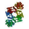
| Deposited unit | 
| ||||||||
|---|---|---|---|---|---|---|---|---|---|
| 1 | 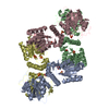
| ||||||||
| 2 | 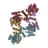
| ||||||||
| 3 | 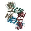
| ||||||||
| Unit cell |
| ||||||||
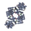
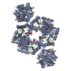
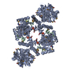
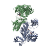
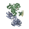
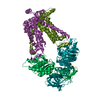
| Details | Biological unit is a homo-tetramer. The ASU contains three such tetramers. |
-Components
| #1: Protein | Mass: 34671.855 Da / Num. of mol.: 12 Source method: isolated from a genetically manipulated source Source: (gene. exp.) Enterobacteria phage T4 (virus) / Genus: T4-like viruses / Species: Enterobacteria phage T4 sensu lato / Gene: pseT / Plasmid: pET28-His10-Smt3 / Species (production host): Escherichia coli / Production host:  References: UniProt: P06855, polynucleotide 5'-hydroxyl-kinase #2: Chemical | ChemComp-MG /   Mass: 24.305 Da / Num. of mol.: 7 / Source method: obtained synthetically / Formula: Mg Mass: 24.305 Da / Num. of mol.: 7 / Source method: obtained synthetically / Formula: Mg#3: Chemical | ChemComp-SO4 /   Mass: 96.063 Da / Num. of mol.: 36 / Source method: obtained synthetically / Formula: SO4 Mass: 96.063 Da / Num. of mol.: 36 / Source method: obtained synthetically / Formula: SO4#4: Chemical | ChemComp-ARS /   Mass: 74.922 Da / Num. of mol.: 11 / Source method: obtained synthetically / Formula: As Mass: 74.922 Da / Num. of mol.: 11 / Source method: obtained synthetically / Formula: As#5: Water | ChemComp-HOH / |  Mass: 18.015 Da / Num. of mol.: 976 / Source method: isolated from a natural source / Formula: H2O Mass: 18.015 Da / Num. of mol.: 976 / Source method: isolated from a natural source / Formula: H2O |
|---|
-Experimental details
-Experiment
| Experiment | Method: X-RAY DIFFRACTION / Number of used crystals: 1 |
|---|
- Sample preparation
| Crystal | Density Matthews: 3.41 Å3/Da / Density % sol: 63.95 % |
|---|---|
| Crystal grow | Temperature: 295 K / Method: vapor diffusion, hanging drop / pH: 6.6 Details: 100mM Sodium cacodylate, 12% PEG-8000, 0.2 M ammonium sulfate, 20 mM urea, 5 mM DTT, pH 6.6, VAPOR DIFFUSION, HANGING DROP, temperature 295K |
-Data collection
| Diffraction | Mean temperature: 130 K |
|---|---|
| Diffraction source | Source: SYNCHROTRON / Site: NSLS  / Beamline: X9A / Wavelength: 0.979 Å / Beamline: X9A / Wavelength: 0.979 Å |
| Detector | Type: MAR CCD 165 mm / Detector: CCD / Date: Aug 15, 2002 Details: Double crystal monochromator with sagitally focusing Si(111) crystals |
| Radiation | Monochromator: Si(111) / Protocol: SINGLE WAVELENGTH / Monochromatic (M) / Laue (L): M / Scattering type: x-ray |
| Radiation wavelength | Wavelength: 0.979 Å / Relative weight: 1 |
| Reflection | Resolution: 2.9→40 Å / Num. all: 126336 / Num. obs: 124693 / % possible obs: 98.7 % / Observed criterion σ(F): -999 / Observed criterion σ(I): -3 / Redundancy: 4.32 % / Biso Wilson estimate: 71.8 Å2 / Rsym value: 0.104 / Net I/σ(I): 12.01 |
| Reflection shell | Resolution: 2.9→2.95 Å / Redundancy: 3.5 % / Mean I/σ(I) obs: 1.57 / Num. unique all: 6245 / Rsym value: 0.535 |
- Processing
| Software |
| ||||||||||||||||||||||||||||||||||||||||||||||||||||||||||||||||||||||||||||||||
|---|---|---|---|---|---|---|---|---|---|---|---|---|---|---|---|---|---|---|---|---|---|---|---|---|---|---|---|---|---|---|---|---|---|---|---|---|---|---|---|---|---|---|---|---|---|---|---|---|---|---|---|---|---|---|---|---|---|---|---|---|---|---|---|---|---|---|---|---|---|---|---|---|---|---|---|---|---|---|---|---|---|
| Refinement | Method to determine structure: MOLECULAR REPLACEMENT Starting model: PDB ID 1LTQ dimer Resolution: 2.9→19.99 Å / Rfactor Rfree error: 0.005 / Data cutoff high absF: 1948026.8 / Data cutoff low absF: 0 / Isotropic thermal model: RESTRAINED / Cross valid method: THROUGHOUT / σ(F): 1 / Stereochemistry target values: Engh & Huber / Details: BULK SOLVENT MODEL USED
| ||||||||||||||||||||||||||||||||||||||||||||||||||||||||||||||||||||||||||||||||
| Solvent computation | Solvent model: FLAT MODEL / Bsol: -0.445649 Å2 / ksol: 0.24 e/Å3 | ||||||||||||||||||||||||||||||||||||||||||||||||||||||||||||||||||||||||||||||||
| Displacement parameters | Biso mean: 60.5 Å2
| ||||||||||||||||||||||||||||||||||||||||||||||||||||||||||||||||||||||||||||||||
| Refine analyze |
| ||||||||||||||||||||||||||||||||||||||||||||||||||||||||||||||||||||||||||||||||
| Refinement step | Cycle: LAST / Resolution: 2.9→19.99 Å
| ||||||||||||||||||||||||||||||||||||||||||||||||||||||||||||||||||||||||||||||||
| Refine LS restraints |
| ||||||||||||||||||||||||||||||||||||||||||||||||||||||||||||||||||||||||||||||||
| Refine LS restraints NCS | NCS model details: CONSTR | ||||||||||||||||||||||||||||||||||||||||||||||||||||||||||||||||||||||||||||||||
| LS refinement shell | Resolution: 2.9→3 Å / Rfactor Rfree error: 0.03 / Total num. of bins used: 10
| ||||||||||||||||||||||||||||||||||||||||||||||||||||||||||||||||||||||||||||||||
| Xplor file |
|
