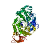
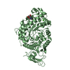
Synthesis of IP2, IP, and Ins in the cytosol / inositol biosynthetic process / inositol-phosphate phosphatase / inositol monophosphate 3-phosphatase activity / inositol monophosphate 4-phosphatase activity / inositol monophosphate 1-phosphatase activity / inositol metabolic process / phosphate-containing compound metabolic process / response to lithium ion / phosphatidylinositol phosphate biosynthetic process ...Synthesis of IP2, IP, and Ins in the cytosol / inositol biosynthetic process / inositol-phosphate phosphatase / inositol monophosphate 3-phosphatase activity / inositol monophosphate 4-phosphatase activity / inositol monophosphate 1-phosphatase activity / inositol metabolic process / phosphate-containing compound metabolic process / response to lithium ion / phosphatidylinositol phosphate biosynthetic process / signal transduction / protein homodimerization activity / metal ion binding / cytosol / cytoplasm Similarity search - Function
Inositol monophosphatase, lithium-sensitive / Inositol monophosphatase / Inositol monophosphatase, conserved site / Inositol monophosphatase family signature 2. / Inositol monophosphatase, metal-binding site / Inositol monophosphatase family signature 1. / Inositol monophosphatase-like / Inositol monophosphatase family / D-Maltodextrin-Binding Protein; domain 2 - #80 / Fructose-1,6-Bisphosphatase, subunit A, domain 1 ...Inositol monophosphatase, lithium-sensitive / Inositol monophosphatase / Inositol monophosphatase, conserved site / Inositol monophosphatase family signature 2. / Inositol monophosphatase, metal-binding site / Inositol monophosphatase family signature 1. / Inositol monophosphatase-like / Inositol monophosphatase family / D-Maltodextrin-Binding Protein; domain 2 - #80 / Fructose-1,6-Bisphosphatase, subunit A, domain 1 / Fructose-1,6-Bisphosphatase; Chain A, domain 1 / D-Maltodextrin-Binding Protein; domain 2 / 2-Layer Sandwich / 3-Layer(aba) Sandwich / Alpha Beta Similarity search - Domain/homology
Resolution: 2.4→2.5 Å / Redundancy: 3.7 % / Mean I/σ(I) obs: 3.2 / Rsym value: 0.433 / % possible all: 99.8
-
Processing
Software
Name
Version
Classification
NB
SHELX
refinement
PDB_EXTRACT
1.701
dataextraction
XDS
datareduction
XSCALE
datascaling
MOLREP
phasing
SHELXL-97
refinement
Refinement
Method to determine structure: MOLECULAR REPLACEMENT / Resolution: 2.4→10 Å / Num. parameters: 30068 / Num. restraintsaints: 31372 / Cross valid method: FREE R / σ(F): 0 / Stereochemistry target values: ENGH & HUBER Details: This is a twinned structure. The twinning operator is (h,k,l) -> (h,-k,-l) and the twinning fraction is 0.5.
In the structure databanks used in Yorodumi, some data are registered as the other names, "COVID-19 virus" and "2019-nCoV". Here are the details of the virus and the list of structure data.
Jan 31, 2019. EMDB accession codes are about to change! (news from PDBe EMDB page)
EMDB accession codes are about to change! (news from PDBe EMDB page)
The allocation of 4 digits for EMDB accession codes will soon come to an end. Whilst these codes will remain in use, new EMDB accession codes will include an additional digit and will expand incrementally as the available range of codes is exhausted. The current 4-digit format prefixed with “EMD-” (i.e. EMD-XXXX) will advance to a 5-digit format (i.e. EMD-XXXXX), and so on. It is currently estimated that the 4-digit codes will be depleted around Spring 2019, at which point the 5-digit format will come into force.
The EM Navigator/Yorodumi systems omit the EMD- prefix.
Related info.:Q: What is EMD? / ID/Accession-code notation in Yorodumi/EM Navigator
Yorodumi is a browser for structure data from EMDB, PDB, SASBDB, etc.
This page is also the successor to EM Navigator detail page, and also detail information page/front-end page for Omokage search.
The word "yorodu" (or yorozu) is an old Japanese word meaning "ten thousand". "mi" (miru) is to see.
Related info.:EMDB / PDB / SASBDB / Comparison of 3 databanks / Yorodumi Search / Aug 31, 2016. New EM Navigator & Yorodumi / Yorodumi Papers / Jmol/JSmol / Function and homology information / Changes in new EM Navigator and Yorodumi
 Movie
Movie Controller


 Open data
 Basic information
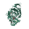
 Components
 Keywords
 Function and homology information
 Authors
 Citation
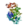
 Structure visualization
 Downloads & links
 Other downloads
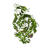

 PDBj
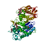
PDBj Assembly

 Sample preparation
 / Beamline: I911-5 / Wavelength: 0.9075 Å
 Processing