 Movie
Movie Controller

[English] 日本語
 Yorodumi
- PDB-2ffg: Novel x-ray structure of the YkuJ protein from Bacillus subtilis.... -
+ Open data

- Basic information
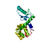
| Entry | Database: PDB / ID: 2ffg | ||||||
|---|---|---|---|---|---|---|---|
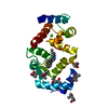
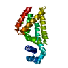
| Title | Novel x-ray structure of the YkuJ protein from Bacillus subtilis. Northeast Structural Genomics target SR360. | ||||||
 Components Components | ykuJ | ||||||
 Keywords Keywords | STRUCTURAL GENOMICS / UNKNOWN FUNCTION / X-Ray SR360 NESG YkuJ / PSI / Protein Structure Initiative / Northeast Structural Genomics Consortium | ||||||
| Function / homology | Protein of unknown function DUF1797 / YkuJ-like / YkuJ-like superfamily / Protein of unknown function (DUF1797) / Signal recognition particle alu RNA binding heterodimer, srp9/1 / 2-Layer Sandwich / Alpha Beta / Uncharacterized protein YkuJ Function and homology information Function and homology information | ||||||
| Biological species |  | ||||||
| Method |  X-RAY DIFFRACTION / SYNCHROTRON / SAD / Resolution: 2.31 Å X-RAY DIFFRACTION / SYNCHROTRON / SAD / Resolution: 2.31 Å | ||||||
 Authors Authors | Kuzin, A.P. / Abashidze, M. / Forouhar, F. / Vorobiev, S.M. / Ho, C.K. / Janjua, H. / Cunningham, K. / Conover, K. / Ma, L.C. / Xiao, R. ...Kuzin, A.P. / Abashidze, M. / Forouhar, F. / Vorobiev, S.M. / Ho, C.K. / Janjua, H. / Cunningham, K. / Conover, K. / Ma, L.C. / Xiao, R. / Acton, T.B. / Montelione, G.T. / Tong, L. / Hunt, J.F. / Northeast Structural Genomics Consortium (NESG) | ||||||
 Citation Citation | Journal: To be Published Title: Novel x-ray structure of the YkuJ protein from Bacillus subtilis. Northeast Structural Genomics target SR360. Authors: Kuzin, A.P. / Abashidze, M. / Forouhar, F. / Vorobiev, S.M. / Ho, C.K. / Janjua, H. / Cunningham, K. / Conover, K. / Ma, L.C. / Xiao, R. / Acton, T.B. / Montelione, G.T. / Tong, L. / Hunt, J. ...Authors: Kuzin, A.P. / Abashidze, M. / Forouhar, F. / Vorobiev, S.M. / Ho, C.K. / Janjua, H. / Cunningham, K. / Conover, K. / Ma, L.C. / Xiao, R. / Acton, T.B. / Montelione, G.T. / Tong, L. / Hunt, J.F. / Northeast Structural Genomics Consortium (NESG) | ||||||
| History |
|
- Structure visualization
| Structure viewer | Molecule: MolmilJmol/JSmol |
|---|
- Downloads & links
-Download
| PDBx/mmCIF format | 2ffg.cif.gz | 41.8 KB | Display | PDBx/mmCIF format |
|---|---|---|---|---|
| PDB format | pdb2ffg.ent.gz | 33.6 KB | Display | PDB format |
| PDBx/mmJSON format | 2ffg.json.gz | Tree view | PDBx/mmJSON format | |
| Others |  Other downloads Other downloads |
-Validation report
| Summary document | 2ffg_validation.pdf.gz | 432.3 KB | Display | wwPDB validaton report |
|---|---|---|---|---|
| Full document | 2ffg_full_validation.pdf.gz | 437.4 KB | Display | |
| Data in XML | 2ffg_validation.xml.gz | 9.6 KB | Display | |
| Data in CIF | 2ffg_validation.cif.gz | 12.2 KB | Display | |
| Arichive directory | https://data.pdbj.org/pub/pdb/validation_reports/ff/2ffgftp://data.pdbj.org/pub/pdb/validation_reports/ff/2ffg | HTTPS FTP |
-Related structure data
| Similar structure data | |
|---|---|
| Other databases |


-Links
 PDBj
PDBj- Assembly
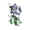
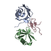
| Deposited unit | 
| ||||||||
|---|---|---|---|---|---|---|---|---|---|
| 1 |
| ||||||||
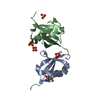
| Unit cell |
|
-Components
| #1: Protein | Mass: 10510.231 Da / Num. of mol.: 2 Source method: isolated from a genetically manipulated source Source: (gene. exp.) #2: Water | ChemComp-HOH / |  Mass: 18.015 Da / Num. of mol.: 67 / Source method: isolated from a natural source / Formula: H2O Mass: 18.015 Da / Num. of mol.: 67 / Source method: isolated from a natural source / Formula: H2O |
|---|
-Experimental details
-Experiment
| Experiment | Method: X-RAY DIFFRACTION / Number of used crystals: 1 |
|---|
- Sample preparation
| Crystal | Density Matthews: 2.25 Å3/Da / Density % sol: 45.44 % |
|---|---|
| Crystal grow | Temperature: 293 K / Method: vapor diffusion, hanging drop / pH: 7.5 Details: 200 mM Ca-acetate, 18% PEG3350, 100 mM Na-Hepes, pH 7.5, VAPOR DIFFUSION, HANGING DROP, temperature 293K |
-Data collection
| Diffraction | Mean temperature: 100 K |
|---|---|
| Diffraction source | Source: SYNCHROTRON / Site: NSLS  / Beamline: X4A / Wavelength: 0.979 Å / Beamline: X4A / Wavelength: 0.979 Å |
| Detector | Type: ADSC QUANTUM 4 / Detector: CCD / Date: Nov 20, 2005 / Details: Mirror |
| Radiation | Protocol: SINGLE WAVELENGTH / Monochromatic (M) / Laue (L): M / Scattering type: x-ray |
| Radiation wavelength | Wavelength: 0.979 Å / Relative weight: 1 |
| Reflection | Resolution: 2.3→30 Å / Num. all: 16561 / Num. obs: 16008 / % possible obs: 99.3 % / Observed criterion σ(F): 2 / Observed criterion σ(I): -3 / Redundancy: 8.3 % / Biso Wilson estimate: 37.6 Å2 / Rmerge(I) obs: 0.06 / Net I/σ(I): 32.5 |
| Reflection shell | Resolution: 2.3→2.38 Å / Rmerge(I) obs: 0.319 / Mean I/σ(I) obs: 5.7 / % possible all: 97.8 |
- Processing
| Software |
| |||||||||||||||||||||||||
|---|---|---|---|---|---|---|---|---|---|---|---|---|---|---|---|---|---|---|---|---|---|---|---|---|---|---|
| Refinement | Method to determine structure: SAD / Resolution: 2.31→19.82 Å / Rfactor Rfree error: 0.01 / Data cutoff high absF: 356919.82 / Data cutoff low absF: 0 / Isotropic thermal model: RESTRAINED / Cross valid method: THROUGHOUT / σ(F): 2 / Stereochemistry target values: Engh & Huber
| |||||||||||||||||||||||||
| Solvent computation | Solvent model: FLAT MODEL / Bsol: 24.3946 Å2 / ksol: 0.292296 e/Å3 | |||||||||||||||||||||||||
| Displacement parameters | Biso mean: 39.5 Å2
| |||||||||||||||||||||||||
| Refine analyze |
| |||||||||||||||||||||||||
| Refinement step | Cycle: LAST / Resolution: 2.31→19.82 Å
| |||||||||||||||||||||||||
| Refine LS restraints |
| |||||||||||||||||||||||||
| LS refinement shell | Resolution: 2.3→2.44 Å / Rfactor Rfree error: 0.024 / Total num. of bins used: 6
| |||||||||||||||||||||||||
| Xplor file |
|
