 Movie
Movie Controller


+ Open data

- Basic information
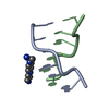
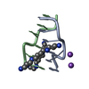
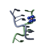
| Entry | Database: PDB / ID: 2f8w | ||||||||||||||||||||
|---|---|---|---|---|---|---|---|---|---|---|---|---|---|---|---|---|---|---|---|---|---|
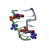
| Title | Crystal structure of d(CACGTG)2 | ||||||||||||||||||||
 Components Components | 5'-D(* Keywords KeywordsDNA / d(CACGTG) / Polyamine / Z-DNA / 1 / 3-propanediamine | Function / homology | 1,3-DIAMINOPROPANE / SPERMINE / DNA |  Function and homology information Function and homology informationMethod |  X-RAY DIFFRACTION / MOLECULAR REPLACEMENT / Resolution: 1.2 Å X-RAY DIFFRACTION / MOLECULAR REPLACEMENT / Resolution: 1.2 Å  Authors AuthorsNarayana, N. / Shamala, N. / Ganesh, K.N. / Viswamitra, M.A. |  CitationJournal: Biochemistry / Year: 2006 CitationJournal: Biochemistry / Year: 2006Title: Interaction between the Z-Type DNA Duplex and 1,3-Propanediamine: Crystal Structure of d(CACGTG)2 at 1.2 A Resolution Authors: Narayana, N. / Shamala, N. / Ganesh, K.N. / Viswamitra, M.A. #1: Journal: Biochemistry / Year: 1991Title: Structure of the pure-spermine form of Z-DNA (magnesium free) at 1-resolution Authors: Egli, M. / Williams, L.D. / Gao, Q. / Rich, A. #2: Journal: Acta Crystallogr.,Sect.D / Year: 2002Title: Structure of d(TGCGCA)2 at 293 K: comparison of the effects of sequence and temperature Authors: Thiyagarajan, S. / Satheesh Kumar, P. / Rajan, S.S. / Gautham, N. #3: Journal: J.Mol.Biol. / Year: 1995Title: Sequence-dependent microheterogeneity of Z-DNA: The crystal and molecular structures of d(CACGCG).d(CGCGTG) and d(CGCACG).d(CGTGCG) Authors: Sadasivan, C. / Gautham, N. #4: Journal: Nucleic Acids Res. / Year: 2004Title: Cobalt hexammine induced tautomeric shift in Z-DNA: the structure of d(CGCGCA)*d(TGCGCG) in two crystal forms Authors: Thiyagarajan, S. / Rajan, S.S. / Gautham, N. #5: Journal: Acta Crystallogr.,Sect.D / Year: 1998Title: Structure of d(TGCGCA)2 and a comparison to other DNA hexamers. Authors: Harper, A. / Brannigan, J.A. / Buck, M. / Hewitt, L. / Lewis, R.J. / Moore, M.H. / Schneider, B. #6: Journal: Nucleic Acids Res. / Year: 1988Title: Structure of d(CACGTG), Z-DNA hexamer containing AT base pairs Authors: Coll, M. / Fita, I. / Lloveras, J. / Subirana, J.A. / Bardella, F. / Huynh-Dinh, T. / Igolen, J. History |
Remark 600 | HETEROGEN Ligand Z5 in the manuscript is labeled as 13D in the coordinate file. | |
- Structure visualization
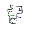
| Structure viewer | Molecule: MolmilJmol/JSmol |
|---|
- Downloads & links
-Download
| PDBx/mmCIF format | 2f8w.cif.gz | 19.4 KB | Display | PDBx/mmCIF format |
|---|---|---|---|---|
| PDB format | pdb2f8w.ent.gz | 11.7 KB | Display | PDB format |
| PDBx/mmJSON format | 2f8w.json.gz | Tree view | PDBx/mmJSON format | |
| Others |  Other downloads Other downloads |
-Validation report
| Arichive directory | https://data.pdbj.org/pub/pdb/validation_reports/f8/2f8wftp://data.pdbj.org/pub/pdb/validation_reports/f8/2f8w | HTTPS FTP |
|---|
-Related structure data
| Related structure data | 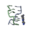 1d48S S: Starting model for refinement |
|---|---|
| Similar structure data |

-Links
 PDBj
PDBj


- Assembly
| Deposited unit | 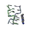
| ||||||||
|---|---|---|---|---|---|---|---|---|---|
| 1 |
| ||||||||
| Unit cell |
| ||||||||
| Details | The biological assembly is a duplex. The coordinates deposited will generate the biological assembly. There is no need for symmetry operations to generate the biological assembly. |
-Components
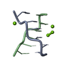
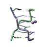
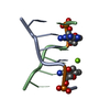
| #1: DNA chain | Mass: 1809.217 Da / Num. of mol.: 2 / Source method: obtained synthetically / Details: synthetic oligonucleotide #2: Chemical | ChemComp-SPM / |   Mass: 202.340 Da / Num. of mol.: 1 / Source method: obtained synthetically / Formula: C10H26N4 Mass: 202.340 Da / Num. of mol.: 1 / Source method: obtained synthetically / Formula: C10H26N4#3: Chemical | ChemComp-13D / |   Mass: 74.125 Da / Num. of mol.: 1 / Source method: obtained synthetically / Formula: C3H10N2 Mass: 74.125 Da / Num. of mol.: 1 / Source method: obtained synthetically / Formula: C3H10N2#4: Water | ChemComp-HOH / |  Mass: 18.015 Da / Num. of mol.: 71 / Source method: isolated from a natural source / Formula: H2O Mass: 18.015 Da / Num. of mol.: 71 / Source method: isolated from a natural source / Formula: H2O |
|---|
-Experimental details
-Experiment
| Experiment | Method: X-RAY DIFFRACTION / Number of used crystals: 1 |
|---|
- Sample preparation
| Crystal | Density Matthews: 1.71 Å3/Da / Density % sol: 28 % | ||||||||||||||||||||||||||||||||||||
|---|---|---|---|---|---|---|---|---|---|---|---|---|---|---|---|---|---|---|---|---|---|---|---|---|---|---|---|---|---|---|---|---|---|---|---|---|---|
| Crystal grow | Temperature: 291 K / Method: vapor diffusion, sitting drop / pH: 6.5 Details: 7.5 mM sodium cacodylate, 2.5 mM SrCl2, 2.5 mM spermine, 50% Isopropanol, pH 6.5, VAPOR DIFFUSION, SITTING DROP, temperature 291K | ||||||||||||||||||||||||||||||||||||
| Components of the solutions |
|
-Data collection
| Diffraction | Mean temperature: 291 K |
|---|---|
| Diffraction source | Source: SEALED TUBE / Type: ENRAF-NONIUS / Wavelength: 1.5418 Å |
| Detector | Type: ENRAF-NONIUS CAD4 / Detector: DIFFRACTOMETER / Date: Feb 4, 1987 |
| Radiation | Monochromator: GRAPHITE / Protocol: SINGLE WAVELENGTH / Monochromatic (M) / Laue (L): M / Scattering type: x-ray |
| Radiation wavelength | Wavelength: 1.5418 Å / Relative weight: 1 |
| Reflection | Resolution: 1.2→50 Å / Num. all: 8124 / Num. obs: 5687 / % possible obs: 70 % / Observed criterion σ(F): 2 / Observed criterion σ(I): 4 / Rsym value: 0.042 |
- Processing
| Software |
| |||||||||||||||||||||||||
|---|---|---|---|---|---|---|---|---|---|---|---|---|---|---|---|---|---|---|---|---|---|---|---|---|---|---|
| Refinement | Method to determine structure: MOLECULAR REPLACEMENT Starting model: PDB entry 1D48 Resolution: 1.2→6 Å / Isotropic thermal model: Isotropic / Cross valid method: THROUGHOUT / σ(F): 2 / σ(I): 4 / Details: X-PLOR
| |||||||||||||||||||||||||
| Displacement parameters | Biso mean: 14.8 Å2 | |||||||||||||||||||||||||
| Refinement step | Cycle: LAST / Resolution: 1.2→6 Å
| |||||||||||||||||||||||||
| Refine LS restraints |
|
