 Movie
Movie Controller

[English] 日本語
 Yorodumi
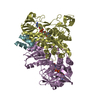
- PDB-2eq7: Crystal structure of lipoamide dehydrogenase from thermus thermop... -
+ Open data

- Basic information
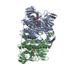
| Entry | Database: PDB / ID: 2eq7 | ||||||
|---|---|---|---|---|---|---|---|
| Title | Crystal structure of lipoamide dehydrogenase from thermus thermophilus HB8 with psbdo | ||||||
 Components Components |
| ||||||
 Keywords Keywords | OXIDOREDUCTASE / PROTEIN-PROTEIN COMPLEX / Structural Genomics / NPPSFA / National Project on Protein Structural and Functional Analyses / RIKEN Structural Genomics/Proteomics Initiative / RSGI | ||||||
| Function / homology |  Function and homology information Function and homology informationdihydrolipoyl dehydrogenase / dihydrolipoyl dehydrogenase (NADH) activity / L-lysine catabolic process to acetyl-CoA via L-saccharopine / dihydrolipoyllysine-residue succinyltransferase / dihydrolipoyllysine-residue succinyltransferase activity / oxoglutarate dehydrogenase complex / 2-oxoglutarate metabolic process / tricarboxylic acid cycle / flavin adenine dinucleotide binding / cytoplasm / cytosol Similarity search - Function | ||||||
| Biological species |   Thermus thermophilus (bacteria) Thermus thermophilus (bacteria) | ||||||
| Method |  X-RAY DIFFRACTION / SYNCHROTRON / MOLECULAR REPLACEMENT / Resolution: 1.8 Å X-RAY DIFFRACTION / SYNCHROTRON / MOLECULAR REPLACEMENT / Resolution: 1.8 Å | ||||||
 Authors Authors | Nakai, T. / Kamiya, N. / RIKEN Structural Genomics/Proteomics Initiative (RSGI) | ||||||
 Citation Citation | Journal: To be Published Title: Crystal structure of lipoamide dehydrogenase from Thermus thermophilus HB8 Authors: Nakai, T. / Kamiya, N. | ||||||
| History |
|
- Structure visualization
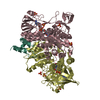
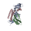
| Structure viewer | Molecule: MolmilJmol/JSmol |
|---|
- Downloads & links
-Download
| PDBx/mmCIF format | 2eq7.cif.gz | 222 KB | Display | PDBx/mmCIF format |
|---|---|---|---|---|
| PDB format | pdb2eq7.ent.gz | 172.9 KB | Display | PDB format |
| PDBx/mmJSON format | 2eq7.json.gz | Tree view | PDBx/mmJSON format | |
| Others |  Other downloads Other downloads |
-Validation report
| Arichive directory | https://data.pdbj.org/pub/pdb/validation_reports/eq/2eq7ftp://data.pdbj.org/pub/pdb/validation_reports/eq/2eq7 | HTTPS FTP |
|---|
-Related structure data
| Related structure data |  2eq6SC 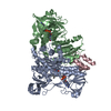 2eq8C 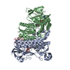 2eq9C S: Starting model for refinement C: citing same article ( |
|---|---|
| Similar structure data | |
| Other databases |


-Links
 PDBj
PDBj


- Assembly
| Deposited unit | 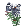
| ||||||||
|---|---|---|---|---|---|---|---|---|---|
| 1 |
| ||||||||
| Unit cell |
|
-Components
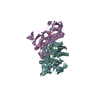
| #1: Protein | Mass: 49127.754 Da / Num. of mol.: 2 Source method: isolated from a genetically manipulated source Source: (gene. exp.) Thermus thermophilus (bacteria) / Strain: HB8 / Gene: TTHA0287 / Plasmid: PET11A / Production host: #2: Protein/peptide | | Mass: 4354.039 Da / Num. of mol.: 1 / Fragment: peripheral subunit binding domain / Source method: obtained synthetically Details: The 40-residue peptide corresponding to a domain of the TTHA0288 protein was synthesized by Greiner References: UniProt: Q5SLK5, dihydrolipoyllysine-residue succinyltransferase #3: Chemical |   Mass: 785.550 Da / Num. of mol.: 2 / Source method: obtained synthetically / Formula: C27H33N9O15P2 / Comment: FAD*YM Mass: 785.550 Da / Num. of mol.: 2 / Source method: obtained synthetically / Formula: C27H33N9O15P2 / Comment: FAD*YM#4: Chemical |   Mass: 663.425 Da / Num. of mol.: 2 / Source method: obtained synthetically / Formula: C21H27N7O14P2 / Comment: NAD*YM Mass: 663.425 Da / Num. of mol.: 2 / Source method: obtained synthetically / Formula: C21H27N7O14P2 / Comment: NAD*YM#5: Water | ChemComp-HOH / |  Mass: 18.015 Da / Num. of mol.: 1159 / Source method: isolated from a natural source / Formula: H2O Mass: 18.015 Da / Num. of mol.: 1159 / Source method: isolated from a natural source / Formula: H2OHas protein modification | Y | |
|---|
-Experimental details
-Experiment
| Experiment | Method: X-RAY DIFFRACTION / Number of used crystals: 1 |
|---|
- Sample preparation
| Crystal | Density Matthews: 2.92 Å3/Da / Density % sol: 57.88 % |
|---|---|
| Crystal grow | Temperature: 291 K / Method: vapor diffusion, hanging drop / pH: 7.5 Details: 45%(v/v) MPD, 200mM NaCl, 10mM NAD+, pH 7.5, VAPOR DIFFUSION, HANGING DROP, temperature 291K |
-Data collection
| Diffraction | Mean temperature: 90 K |
|---|---|
| Diffraction source | Source: SYNCHROTRON / Site: SPring-8  / Beamline: BL44B2 / Wavelength: 1 Å / Beamline: BL44B2 / Wavelength: 1 Å |
| Detector | Type: ADSC QUANTUM 210 / Detector: CCD / Date: Jul 15, 2006 |
| Radiation | Monochromator: SI(111) / Protocol: SINGLE WAVELENGTH / Monochromatic (M) / Laue (L): M / Scattering type: x-ray |
| Radiation wavelength | Wavelength: 1 Å / Relative weight: 1 |
| Reflection | Resolution: 1.8→50 Å / Num. obs: 111571 / % possible obs: 99.8 % / Observed criterion σ(I): 0 / Redundancy: 7.1 % / Biso Wilson estimate: 14.6 Å2 / Rmerge(I) obs: 0.075 / Net I/σ(I): 25.6 |
| Reflection shell | Resolution: 1.8→1.86 Å / Redundancy: 6.6 % / Rmerge(I) obs: 0.413 / Mean I/σ(I) obs: 3.6 / Num. unique all: 10985 / % possible all: 99.6 |
- Processing
| Software |
| ||||||||||||||||||||||||||||||||||||||||||||||||||||||||||||||||||||||||||||||||
|---|---|---|---|---|---|---|---|---|---|---|---|---|---|---|---|---|---|---|---|---|---|---|---|---|---|---|---|---|---|---|---|---|---|---|---|---|---|---|---|---|---|---|---|---|---|---|---|---|---|---|---|---|---|---|---|---|---|---|---|---|---|---|---|---|---|---|---|---|---|---|---|---|---|---|---|---|---|---|---|---|---|
| Refinement | Method to determine structure: MOLECULAR REPLACEMENT Starting model: PDB ENTRY 2EQ6 Resolution: 1.8→39.01 Å / Rfactor Rfree error: 0.003 / Data cutoff high absF: 2723055.64 / Data cutoff low absF: 0 / Isotropic thermal model: RESTRAINED / Cross valid method: THROUGHOUT / σ(F): 0 / Stereochemistry target values: Engh & Huber
| ||||||||||||||||||||||||||||||||||||||||||||||||||||||||||||||||||||||||||||||||
| Solvent computation | Solvent model: FLAT MODEL / Bsol: 72.0469 Å2 / ksol: 0.345653 e/Å3 | ||||||||||||||||||||||||||||||||||||||||||||||||||||||||||||||||||||||||||||||||
| Displacement parameters | Biso mean: 23.2 Å2
| ||||||||||||||||||||||||||||||||||||||||||||||||||||||||||||||||||||||||||||||||
| Refine analyze |
| ||||||||||||||||||||||||||||||||||||||||||||||||||||||||||||||||||||||||||||||||
| Refinement step | Cycle: LAST / Resolution: 1.8→39.01 Å
| ||||||||||||||||||||||||||||||||||||||||||||||||||||||||||||||||||||||||||||||||
| Refine LS restraints |
| ||||||||||||||||||||||||||||||||||||||||||||||||||||||||||||||||||||||||||||||||
| LS refinement shell | Resolution: 1.8→1.91 Å / Rfactor Rfree error: 0.009 / Total num. of bins used: 6
| ||||||||||||||||||||||||||||||||||||||||||||||||||||||||||||||||||||||||||||||||
| Xplor file |
|
