 Movie
Movie Controller

[English] 日本語
 Yorodumi
- PDB-2dze: Crystal structure of histone chaperone Asf1 in complex with a C-t... -
+ Open data

- Basic information
| Entry | Database: PDB / ID: 2dze | ||||||
|---|---|---|---|---|---|---|---|
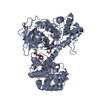
| Title | Crystal structure of histone chaperone Asf1 in complex with a C-terminus of histone H3 | ||||||
 Components Components |
| ||||||
 Keywords Keywords | CHAPERONE / Immunoglobulin fold / Structural Genomics / NPPSFA / National Project on Protein Structural and Functional Analyses / RIKEN Structural Genomics/Proteomics Initiative / RSGI | ||||||
| Function / homology |  Function and homology information Function and homology informationCondensation of Prophase Chromosomes / : / : / Assembly of the ORC complex at the origin of replication / Oxidative Stress Induced Senescence / : / Factors involved in megakaryocyte development and platelet production / nucleolar peripheral inclusion body / Estrogen-dependent gene expression / subtelomeric heterochromatin ...Condensation of Prophase Chromosomes / : / : / Assembly of the ORC complex at the origin of replication / Oxidative Stress Induced Senescence / : / Factors involved in megakaryocyte development and platelet production / nucleolar peripheral inclusion body / Estrogen-dependent gene expression / subtelomeric heterochromatin / RNA Polymerase I Promoter Escape / Transcriptional regulation by small RNAs / mating-type region heterochromatin / heterochromatin boundary formation / H3-H4 histone complex chaperone activity / mitotic sister chromatid biorientation / histone chaperone activity / DNA replication-dependent chromatin assembly / pericentric heterochromatin formation / chromatin-protein adaptor activity / nucleosome disassembly / pericentric heterochromatin / heterochromatin / transcription initiation-coupled chromatin remodeling / structural constituent of chromatin / heterochromatin formation / nucleosome / nucleosome assembly / chromatin organization / histone binding / protein heterodimerization activity / DNA damage response / chromatin / DNA binding / nucleus Similarity search - Function | ||||||
| Biological species |  | ||||||
| Method |  X-RAY DIFFRACTION / SYNCHROTRON / MOLECULAR REPLACEMENT / Resolution: 1.8 Å X-RAY DIFFRACTION / SYNCHROTRON / MOLECULAR REPLACEMENT / Resolution: 1.8 Å | ||||||
 Authors Authors | Padmanabhan, B. / Yokoyama, S. / RIKEN Structural Genomics/Proteomics Initiative (RSGI) | ||||||
 Citation Citation | Journal: To be Published Title: Crystal structure of histone chaperone Asf1 in complex with a C-terminus of histone H3 Authors: Padmanabhan, B. / Yokoyama, S. | ||||||
| History |
|
- Structure visualization
| Structure viewer | Molecule: MolmilJmol/JSmol |
|---|
- Downloads & links
-Download
| PDBx/mmCIF format | 2dze.cif.gz | 83.3 KB | Display | PDBx/mmCIF format |
|---|---|---|---|---|
| PDB format | pdb2dze.ent.gz | 61.7 KB | Display | PDB format |
| PDBx/mmJSON format | 2dze.json.gz | Tree view | PDBx/mmJSON format | |
| Others |  Other downloads Other downloads |
-Validation report
| Arichive directory | https://data.pdbj.org/pub/pdb/validation_reports/dz/2dzeftp://data.pdbj.org/pub/pdb/validation_reports/dz/2dze | HTTPS FTP |
|---|
-Related structure data
| Related structure data |  2cu9S S: Starting model for refinement |
|---|---|
| Similar structure data | |
| Other databases |


-Links
 PDBj
PDBj


- Assembly
| Deposited unit | 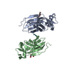
| |||||||||||||||||||||||||||||||||||||||||||||||||||||||||||||||||||||||||||||||||||||||||||||||||||||||||||||||||||||
|---|---|---|---|---|---|---|---|---|---|---|---|---|---|---|---|---|---|---|---|---|---|---|---|---|---|---|---|---|---|---|---|---|---|---|---|---|---|---|---|---|---|---|---|---|---|---|---|---|---|---|---|---|---|---|---|---|---|---|---|---|---|---|---|---|---|---|---|---|---|---|---|---|---|---|---|---|---|---|---|---|---|---|---|---|---|---|---|---|---|---|---|---|---|---|---|---|---|---|---|---|---|---|---|---|---|---|---|---|---|---|---|---|---|---|---|---|---|---|
| 1 |
| |||||||||||||||||||||||||||||||||||||||||||||||||||||||||||||||||||||||||||||||||||||||||||||||||||||||||||||||||||||
| Unit cell |
| |||||||||||||||||||||||||||||||||||||||||||||||||||||||||||||||||||||||||||||||||||||||||||||||||||||||||||||||||||||
| Noncrystallographic symmetry (NCS) | NCS domain:
NCS domain segments: Ens-ID: 1 / Refine code: 5
|
-Components
| #1: Protein | Mass: 18347.934 Da / Num. of mol.: 2 / Fragment: N-termianl region, residues 1-162 Source method: isolated from a genetically manipulated source Source: (gene. exp.) Gene: cia1 / Plasmid: pET15b / Production host:  #2: Protein/peptide | | Mass: 1290.582 Da / Num. of mol.: 1 / Fragment: C-terminal H3 peptide / Source method: obtained synthetically / Details: Synthetic peptide / References: UniProt: P09988 #3: Chemical |   Mass: 150.173 Da / Num. of mol.: 2 / Source method: obtained synthetically / Formula: C6H14O4 Mass: 150.173 Da / Num. of mol.: 2 / Source method: obtained synthetically / Formula: C6H14O4#4: Water | ChemComp-HOH / |  Mass: 18.015 Da / Num. of mol.: 219 / Source method: isolated from a natural source / Formula: H2O Mass: 18.015 Da / Num. of mol.: 219 / Source method: isolated from a natural source / Formula: H2O |
|---|
-Experimental details
-Experiment
| Experiment | Method: X-RAY DIFFRACTION / Number of used crystals: 1 |
|---|
- Sample preparation
| Crystal | Density Matthews: 3.02 Å3/Da / Density % sol: 59.33 % |
|---|---|
| Crystal grow | Temperature: 293 K / Method: vapor diffusion, sitting drop / pH: 8 Details: PEG6000, AS, pH 8.0, VAPOR DIFFUSION, SITTING DROP, temperature 293K |
-Data collection
| Diffraction | Mean temperature: 120 K |
|---|---|
| Diffraction source | Source: SYNCHROTRON / Site: SPring-8  / Beamline: BL26B1 / Wavelength: 1 Å / Beamline: BL26B1 / Wavelength: 1 Å |
| Detector | Type: RIGAKU JUPITER 210 / Detector: CCD / Date: Jul 14, 2005 |
| Radiation | Protocol: SINGLE WAVELENGTH / Monochromatic (M) / Laue (L): M / Scattering type: x-ray |
| Radiation wavelength | Wavelength: 1 Å / Relative weight: 1 |
| Reflection | Resolution: 1.8→50 Å / Num. all: 41187 / Num. obs: 38738 / % possible obs: 94.1 % / Observed criterion σ(I): -3 / Redundancy: 2.3 % / Rmerge(I) obs: 0.056 |
| Reflection shell | Resolution: 1.8→1.86 Å / Redundancy: 2 % / Rmerge(I) obs: 0.191 / Num. unique all: 3746 / % possible all: 91.4 |
- Processing
| Software |
| ||||||||||||||||||||||||||||||||||||||||||||||||||||||||||||||||||||||
|---|---|---|---|---|---|---|---|---|---|---|---|---|---|---|---|---|---|---|---|---|---|---|---|---|---|---|---|---|---|---|---|---|---|---|---|---|---|---|---|---|---|---|---|---|---|---|---|---|---|---|---|---|---|---|---|---|---|---|---|---|---|---|---|---|---|---|---|---|---|---|---|
| Refinement | Method to determine structure: MOLECULAR REPLACEMENT Starting model: PDB Entry 2CU9 Resolution: 1.8→50 Å / Cor.coef. Fo:Fc: 0.939 / Cor.coef. Fo:Fc free: 0.932 / SU B: 2.63 / SU ML: 0.083 / Cross valid method: THROUGHOUT / σ(F): 0 / ESU R: 0.134 / ESU R Free: 0.127 / Stereochemistry target values: MAXIMUM LIKELIHOOD
| ||||||||||||||||||||||||||||||||||||||||||||||||||||||||||||||||||||||
| Solvent computation | Ion probe radii: 0.8 Å / Shrinkage radii: 0.8 Å / VDW probe radii: 1.4 Å / Solvent model: BABINET MODEL WITH MASK | ||||||||||||||||||||||||||||||||||||||||||||||||||||||||||||||||||||||
| Displacement parameters | Biso mean: 27.12 Å2
| ||||||||||||||||||||||||||||||||||||||||||||||||||||||||||||||||||||||
| Refinement step | Cycle: LAST / Resolution: 1.8→50 Å
| ||||||||||||||||||||||||||||||||||||||||||||||||||||||||||||||||||||||
| Refine LS restraints |
| ||||||||||||||||||||||||||||||||||||||||||||||||||||||||||||||||||||||
| Refine LS restraints NCS | Dom-ID: 1 / Auth asym-ID: A / Ens-ID: 1 / Refine-ID: X-RAY DIFFRACTION
| ||||||||||||||||||||||||||||||||||||||||||||||||||||||||||||||||||||||
| LS refinement shell | Resolution: 1.799→1.846 Å / Total num. of bins used: 20 /
|
