 Movie
Movie Controller

[English] 日本語
 Yorodumi
- PDB-2co7: Salmonella enterica SafA pilin in complex with the SafB chaperone... -
+ Open data

- Basic information
| Entry | Database: PDB / ID: 2co7 | ||||||
|---|---|---|---|---|---|---|---|
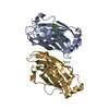
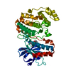
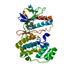
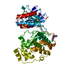
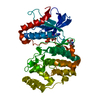
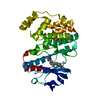
| Title | Salmonella enterica SafA pilin in complex with the SafB chaperone (Type II) | ||||||
 Components Components |
| ||||||
 Keywords Keywords | FIBRIL PROTEIN / PILUS SUBUNIT / CHAPERONE / ADHESION / STRAND COMPLEMENTATION / PATHOGENESIS | ||||||
| Function / homology |  Function and homology information Function and homology informationprotein folding chaperone / cell wall organization / outer membrane-bounded periplasmic space Similarity search - Function | ||||||
| Biological species |  SALMONELLA TYPHIMURIUM (bacteria) SALMONELLA TYPHIMURIUM (bacteria) | ||||||
| Method |  X-RAY DIFFRACTION / SYNCHROTRON / MOLECULAR REPLACEMENT / Resolution: 1.8 Å X-RAY DIFFRACTION / SYNCHROTRON / MOLECULAR REPLACEMENT / Resolution: 1.8 Å | ||||||
 Authors Authors | Remaut, H. / Rose, R.J. / Hannan, T.J. / Hultgren, S.J. / Radford, S.E. / Ashcroft, A.E. / Waksman, G. | ||||||
 Citation Citation | Journal: Mol.Cell / Year: 2006 Title: Donor-Strand Exchange in Chaperone-Assisted Pilus Assembly Proceeds Through a Concerted Beta-Strand Displacement Mechanism Authors: Remaut, H. / Rose, R.J. / Hannan, T.J. / Hultgren, S.J. / Radford, S.E. / Ashcroft, A.E. / Waksman, G. | ||||||
| History |
| ||||||
| Remark 700 | SHEET THE SHEET STRUCTURE OF THIS MOLECULE IS BIFURCATED. IN ORDER TO REPRESENT THIS FEATURE IN ... SHEET THE SHEET STRUCTURE OF THIS MOLECULE IS BIFURCATED. IN ORDER TO REPRESENT THIS FEATURE IN THE SHEET RECORDS BELOW, TWO SHEETS ARE DEFINED. |
- Structure visualization
| Structure viewer | Molecule: MolmilJmol/JSmol |
|---|
- Downloads & links
-Download
| PDBx/mmCIF format | 2co7.cif.gz | 80.9 KB | Display | PDBx/mmCIF format |
|---|---|---|---|---|
| PDB format | pdb2co7.ent.gz | 59.9 KB | Display | PDB format |
| PDBx/mmJSON format | 2co7.json.gz | Tree view | PDBx/mmJSON format | |
| Others |  Other downloads Other downloads |
-Validation report
| Arichive directory | https://data.pdbj.org/pub/pdb/validation_reports/co/2co7ftp://data.pdbj.org/pub/pdb/validation_reports/co/2co7 | HTTPS FTP |
|---|
-Related structure data
| Related structure data | 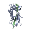 2cnyC  2cnzC 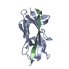 2co1C  2co2C  2co3C 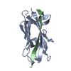 2co4C  2co6C  1l4iS C: citing same article ( S: Starting model for refinement |
|---|---|
| Similar structure data |


-Links
 PDBj
PDBj


- Assembly
| Deposited unit | 
| |||||||||
|---|---|---|---|---|---|---|---|---|---|---|
| 1 |
| |||||||||
| Unit cell |
| |||||||||
| Components on special symmetry positions |
| |||||||||
| Details | FOR THE HETERO-ASSEMBLY DESCRIBED BY REMARK 350THE N-TERMINAL EXTENSION PEPTIDE OF ONE SUBUNIT, CHAIN B,INSERTS INTO THE FOLD OF ANOTHER, CHAIN A. THIS INTERACTIONIS REPETED IN THE POLYMER, AN N-TERMINAL EXTENSION OFMOLECULE C WOULD INSERT IN TO THE SUBUNIT OF MOLECULE B , DINTO C ETC |
-Components
| #1: Protein | Mass: 13361.907 Da / Num. of mol.: 1 / Fragment: CORE PILIN DOMAIN, NTE DELETED, RESIDUES 46-170 / Mutation: YES Source method: isolated from a genetically manipulated source Source: (gene. exp.) SALMONELLA TYPHIMURIUM (bacteria) / Strain: LT2 / Plasmid: PTRC99A / Production host: | ||||||
|---|---|---|---|---|---|---|---|
| #2: Protein | Mass: 24023.285 Da / Num. of mol.: 1 Source method: isolated from a genetically manipulated source Source: (gene. exp.) SALMONELLA TYPHIMURIUM (bacteria) / Strain: LT2 / Plasmid: PTRC99A / Production host: | ||||||
| #3: Chemical |   Mass: 96.063 Da / Num. of mol.: 2 / Source method: obtained synthetically / Formula: SO4 Mass: 96.063 Da / Num. of mol.: 2 / Source method: obtained synthetically / Formula: SO4#4: Water | ChemComp-HOH / |  Mass: 18.015 Da / Num. of mol.: 274 / Source method: isolated from a natural source / Formula: H2O Mass: 18.015 Da / Num. of mol.: 274 / Source method: isolated from a natural source / Formula: H2OHas protein modification | Y | Sequence details | RESIDUES IN THE NTD OF CHAIN A WERE DELETED IN THE SAMPLE USED IN THE EXPERIMENT | |
-Experimental details
-Experiment
| Experiment | Method: X-RAY DIFFRACTION / Number of used crystals: 1 |
|---|
- Sample preparation
| Crystal | Density Matthews: 2.22 Å3/Da / Density % sol: 44.2 % |
|---|---|
| Crystal grow | Method: vapor diffusion, sitting drop Details: 20% PEG 4000, 200 MM AMMONIUM ACETATE, 10% PEG 1000 |
-Data collection
| Diffraction | Mean temperature: 100 K |
|---|---|
| Diffraction source | Source: SYNCHROTRON / Site: ESRF  / Beamline: ID14-1 / Wavelength: 0.9792 / Beamline: ID14-1 / Wavelength: 0.9792 |
| Detector | Type: ADSC CCD / Detector: CCD |
| Radiation | Protocol: SINGLE WAVELENGTH / Monochromatic (M) / Laue (L): M / Scattering type: x-ray |
| Radiation wavelength | Wavelength: 0.9792 Å / Relative weight: 1 |
| Reflection | Resolution: 1.8→25 Å / Num. obs: 27689 / % possible obs: 91.5 % / Observed criterion σ(I): 0 / Redundancy: 4.1 % / Biso Wilson estimate: 18.3 Å2 / Rmerge(I) obs: 0.066 / Net I/σ(I): 17.5 |
| Reflection shell | Resolution: 1.8→1.9 Å / Redundancy: 3.7 % / Rmerge(I) obs: 0.28 / Mean I/σ(I) obs: 4.3 / % possible all: 89.7 |
- Processing
| Software |
| ||||||||||||||||||||||||||||||||||||||||||||||||||||||||||||||||||||||||||||||||||||||||||||||||||||||||||||||||||||||||||||||||||||||||||||||||||||||||||||||||||||||||||||||||||||||
|---|---|---|---|---|---|---|---|---|---|---|---|---|---|---|---|---|---|---|---|---|---|---|---|---|---|---|---|---|---|---|---|---|---|---|---|---|---|---|---|---|---|---|---|---|---|---|---|---|---|---|---|---|---|---|---|---|---|---|---|---|---|---|---|---|---|---|---|---|---|---|---|---|---|---|---|---|---|---|---|---|---|---|---|---|---|---|---|---|---|---|---|---|---|---|---|---|---|---|---|---|---|---|---|---|---|---|---|---|---|---|---|---|---|---|---|---|---|---|---|---|---|---|---|---|---|---|---|---|---|---|---|---|---|---|---|---|---|---|---|---|---|---|---|---|---|---|---|---|---|---|---|---|---|---|---|---|---|---|---|---|---|---|---|---|---|---|---|---|---|---|---|---|---|---|---|---|---|---|---|---|---|---|---|
| Refinement | Method to determine structure: MOLECULAR REPLACEMENT Starting model: PDB ENTRY 1L4I Resolution: 1.8→20 Å / Cor.coef. Fo:Fc: 0.946 / Cor.coef. Fo:Fc free: 0.925 / SU B: 4.695 / SU ML: 0.083 / TLS residual ADP flag: LIKELY RESIDUAL / Cross valid method: THROUGHOUT / ESU R: 0.149 / ESU R Free: 0.138 / Stereochemistry target values: MAXIMUM LIKELIHOOD / Details: HYDROGENS HAVE BEEN ADDED IN THE RIDING POSITIONS.
| ||||||||||||||||||||||||||||||||||||||||||||||||||||||||||||||||||||||||||||||||||||||||||||||||||||||||||||||||||||||||||||||||||||||||||||||||||||||||||||||||||||||||||||||||||||||
| Solvent computation | Ion probe radii: 0.8 Å / Shrinkage radii: 0.8 Å / VDW probe radii: 1.4 Å / Solvent model: MASK | ||||||||||||||||||||||||||||||||||||||||||||||||||||||||||||||||||||||||||||||||||||||||||||||||||||||||||||||||||||||||||||||||||||||||||||||||||||||||||||||||||||||||||||||||||||||
| Displacement parameters | Biso mean: 22.98 Å2
| ||||||||||||||||||||||||||||||||||||||||||||||||||||||||||||||||||||||||||||||||||||||||||||||||||||||||||||||||||||||||||||||||||||||||||||||||||||||||||||||||||||||||||||||||||||||
| Refinement step | Cycle: LAST / Resolution: 1.8→20 Å
| ||||||||||||||||||||||||||||||||||||||||||||||||||||||||||||||||||||||||||||||||||||||||||||||||||||||||||||||||||||||||||||||||||||||||||||||||||||||||||||||||||||||||||||||||||||||
| Refine LS restraints |
|