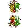
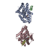
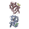
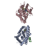
Entry Database : PDB / ID : 2cgyTitle STRUCTURE OF HELIX POMATIA AGGLUTININ WITH FORSMANN ANTIGEN AGGLUTININ Keywords / / Function / homology Function Domain/homology Component
/ / / / / / / / / / / / / / Biological species HELIX POMATIA (Roman snail)Method / / / Resolution : 2.8 Å Authors Sanchez, J.-F. / Lescar, J. / Audfray, A. / Gautier, C. / Chazalet, V. / Gagnon, J. / Breton, C. / Imberty, A. / Mitchell, E.P. Journal : Glycobiology / Year : 2007Title : Structural Basis for Recognition of Breast and Colon Cancer Epitopes Tn Antigen and Forssman Disaccharide by Helix Pomatia Lectin.Authors : Lescar, J. / Sanchez, J.-F. / Audfray, A. / Coll, J. / Breton, C. / Mitchell, E.P. / Imberty, A. History Deposition Mar 10, 2006 Deposition site / Processing site Revision 1.0 Mar 13, 2007 Provider / Type Revision 1.1 Sep 28, 2011 Group Database references / Derived calculations ... Database references / Derived calculations / Non-polymer description / Other / Structure summary / Version format compliance Revision 1.2 May 8, 2019 Group / Experimental preparation / OtherCategory database_PDB_rev / database_PDB_rev_record ... database_PDB_rev / database_PDB_rev_record / exptl_crystal_grow / pdbx_database_proc / pdbx_database_status Item / _pdbx_database_status.recvd_author_approvalRevision 2.0 Jul 29, 2020 Group Atomic model / Data collection ... Atomic model / Data collection / Derived calculations / Non-polymer description / Structure summary Category atom_site / chem_comp ... atom_site / chem_comp / entity / entity_name_com / pdbx_branch_scheme / pdbx_chem_comp_identifier / pdbx_entity_branch / pdbx_entity_branch_descriptor / pdbx_entity_branch_link / pdbx_entity_branch_list / pdbx_entity_nonpoly / pdbx_molecule_features / pdbx_nonpoly_scheme / struct_conn / struct_site / struct_site_gen Item _atom_site.B_iso_or_equiv / _atom_site.Cartn_x ... _atom_site.B_iso_or_equiv / _atom_site.Cartn_x / _atom_site.Cartn_y / _atom_site.Cartn_z / _atom_site.auth_asym_id / _atom_site.auth_atom_id / _atom_site.auth_comp_id / _atom_site.auth_seq_id / _atom_site.label_asym_id / _atom_site.label_atom_id / _atom_site.label_comp_id / _atom_site.label_entity_id / _atom_site.type_symbol / _chem_comp.formula / _chem_comp.formula_weight / _chem_comp.id / _chem_comp.mon_nstd_flag / _chem_comp.name / _chem_comp.pdbx_synonyms / _chem_comp.type / _entity.formula_weight / _entity.pdbx_description / _entity.src_method / _entity.type / _struct_conn.conn_type_id / _struct_conn.id / _struct_conn.pdbx_dist_value / _struct_conn.pdbx_leaving_atom_flag / _struct_conn.pdbx_role / _struct_conn.pdbx_value_order / _struct_conn.ptnr1_auth_asym_id / _struct_conn.ptnr1_auth_comp_id / _struct_conn.ptnr1_auth_seq_id / _struct_conn.ptnr1_label_asym_id / _struct_conn.ptnr1_label_atom_id / _struct_conn.ptnr1_label_comp_id / _struct_conn.ptnr1_label_seq_id / _struct_conn.ptnr2_auth_asym_id / _struct_conn.ptnr2_auth_comp_id / _struct_conn.ptnr2_auth_seq_id / _struct_conn.ptnr2_label_asym_id / _struct_conn.ptnr2_label_atom_id / _struct_conn.ptnr2_label_comp_id / _struct_conn.ptnr2_label_seq_id / _struct_conn.ptnr2_symmetry Description / Provider / Type Revision 2.1 Dec 13, 2023 Group Data collection / Database references ... Data collection / Database references / Refinement description / Structure summary Category chem_comp / chem_comp_atom ... chem_comp / chem_comp_atom / chem_comp_bond / database_2 / pdbx_initial_refinement_model Item / _database_2.pdbx_DOI / _database_2.pdbx_database_accessionRevision 2.2 Nov 6, 2024 Group / Category / pdbx_modification_feature
Show all Show less Remark 650 HELIX DETERMINATION METHOD: AUTHOR PROVIDED. Remark 700 SHEET DETERMINATION METHOD: AUTHOR PROVIDED.
 Movie
Movie Controller


 Open data
 Basic information
 Components
 Keywords
 Function and homology information
 HELIX POMATIA (Roman snail)
 X-RAY DIFFRACTION /
  Authors
 Citation
 Structure visualization
 Downloads & links
 Other downloads
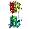


 PDBj
PDBj
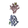
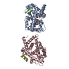
 Assembly
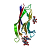


 Type: D-saccharide, beta linking / Mass: 221.208 Da / Num. of mol.: 1
 Mass: 18.015 Da / Num. of mol.: 1 / Source method: isolated from a natural source / Formula: H2O
 Sample preparation
 / Beamline: ID14-2 / Wavelength: 0.933
  Processing