Mass: 18.015 Da / Num. of mol.: 225 / Source method: isolated from a natural source / Formula: H2O
Sequence details
RESIDUE NUMBER BEFORE 1 IS T7 TAG. THE T7 TAGS IN BOTH CHAIN A AND B ARE DISORDERED, AND NOT ...RESIDUE NUMBER BEFORE 1 IS T7 TAG. THE T7 TAGS IN BOTH CHAIN A AND B ARE DISORDERED, AND NOT DETERMINED IN THE MODEL.
-
Experimental details
-
Experiment
Experiment
Method: X-RAY DIFFRACTION
-
Sample preparation
Crystal
Density Matthews: 2.3 Å3/Da / Density % sol: 46.4 %
Protocol: SINGLE WAVELENGTH / Monochromatic (M) / Laue (L): M / Scattering type: x-ray
Radiation wavelength
Wavelength: 1.12714 Å / Relative weight: 1
Reflection
Resolution: 2.4→29.44 Å / Num. obs: 12691 / % possible obs: 99.2 % / Observed criterion σ(I): 2 / Redundancy: 8.6 % / Biso Wilson estimate: 30.7 Å2 / Rmerge(I) obs: 0.05 / Net I/σ(I): 45.1
Reflection shell
Resolution: 2.4→2.49 Å / Redundancy: 6.9 % / Rmerge(I) obs: 0.53 / Mean I/σ(I) obs: 3.8 / % possible all: 98.1
-
Processing
Software
Name
Version
Classification
CNS
1.1
refinement
HKL-2000
datareduction
HKL-2000
datascaling
SOLVE
phasing
Refinement
Method to determine structure: MAD / Resolution: 2.4→29.44 Å / Rfactor Rfree error: 0.007 / Data cutoff high absF: 577606.57 / Isotropic thermal model: RESTRAINED / Cross valid method: THROUGHOUT / σ(F): 2 Details: RESIDUE 96-98 IN CHAIN B ARE DISORDERED, AND NOT DETERMINED IN THE MODEL.
In the structure databanks used in Yorodumi, some data are registered as the other names, "COVID-19 virus" and "2019-nCoV". Here are the details of the virus and the list of structure data.
Jan 31, 2019. EMDB accession codes are about to change! (news from PDBe EMDB page)
EMDB accession codes are about to change! (news from PDBe EMDB page)
The allocation of 4 digits for EMDB accession codes will soon come to an end. Whilst these codes will remain in use, new EMDB accession codes will include an additional digit and will expand incrementally as the available range of codes is exhausted. The current 4-digit format prefixed with “EMD-” (i.e. EMD-XXXX) will advance to a 5-digit format (i.e. EMD-XXXXX), and so on. It is currently estimated that the 4-digit codes will be depleted around Spring 2019, at which point the 5-digit format will come into force.
The EM Navigator/Yorodumi systems omit the EMD- prefix.
Related info.:Q: What is EMD? / ID/Accession-code notation in Yorodumi/EM Navigator
Yorodumi is a browser for structure data from EMDB, PDB, SASBDB, etc.
This page is also the successor to EM Navigator detail page, and also detail information page/front-end page for Omokage search.
The word "yorodu" (or yorozu) is an old Japanese word meaning "ten thousand". "mi" (miru) is to see.
Related info.:EMDB / PDB / SASBDB / Comparison of 3 databanks / Yorodumi Search / Aug 31, 2016. New EM Navigator & Yorodumi / Yorodumi Papers / Jmol/JSmol / Function and homology information / Changes in new EM Navigator and Yorodumi
 Movie
Movie Controller

 Yorodumi
 Open data
 Basic information
 Components
 Keywords
 Function and homology information
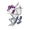
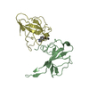
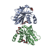
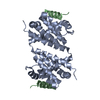
 HOMO SAPIENS (human)
 X-RAY DIFFRACTION /
  Authors
 Citation
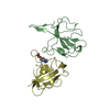
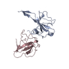
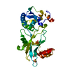
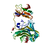
 Structure visualization
 Downloads & links
 Other downloads


 PDBj
PDBj


 Assembly
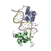


 Mass: 24.305 Da / Num. of mol.: 2 / Source method: obtained synthetically / Formula: Mg
 Mass: 18.015 Da / Num. of mol.: 225 / Source method: isolated from a natural source / Formula: H2O
 Sample preparation
 / Beamline: BL17B2 / Wavelength: 1.12714
  Processing