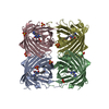
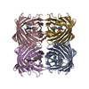
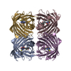
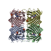
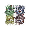
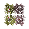
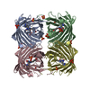
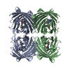
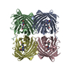
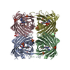
- PDB-2btj: Fluorescent Protein EosFP - red form -
+
Open data
ID or keywords:
Loading...
-
Basic information
Entry
Database: PDB / ID: 2btj
Title
Fluorescent Protein EosFP - red form
Components
(Green to red photoconvertible GFP-like protein EosFP) x 2
Keywords
FLUORESCENT PROTEIN / PHOTO-INDUCED PROTEIN CLEAVAGE / GREEN-TO-RED CONVERSION / LUMINESCENT PROTEIN
Function / homology
Green fluorescent protein, GFP / Green fluorescent protein-related / Green fluorescent protein / Green fluorescent protein / bioluminescence / generation of precursor metabolites and energy / Green to red photoconvertible GFP-like protein EosFP
Function and homology information
Biological species
Lobophyllia hemprichii (invertebrata)
Method
X-RAY DIFFRACTION / MOLECULAR REPLACEMENT / Resolution: 2 Å
SHEET DETERMINATION METHOD: DSSP THE SHEETS PRESENTED AS "AA", "BA", "CA" AND "DA" IN EACH CHAIN ... SHEET DETERMINATION METHOD: DSSP THE SHEETS PRESENTED AS "AA", "BA", "CA" AND "DA" IN EACH CHAIN ON SHEET RECORDS BELOW IS ACTUALLY AN 11-STRANDED BARREL THIS IS REPRESENTED BY A 12-STRANDED SHEET IN WHICH THE FIRST AND LAST STRANDS ARE IDENTICAL.
aa: Green to red photoconvertible GFP-like protein EosFP A: Green to red photoconvertible GFP-like protein EosFP bb: Green to red photoconvertible GFP-like protein EosFP B: Green to red photoconvertible GFP-like protein EosFP cc: Green to red photoconvertible GFP-like protein EosFP C: Green to red photoconvertible GFP-like protein EosFP dd: Green to red photoconvertible GFP-like protein EosFP D: Green to red photoconvertible GFP-like protein EosFP
Mass: 6553.457 Da / Num. of mol.: 4 Source method: isolated from a genetically manipulated source Details: 2-((1E)-2-(5-IMIDAZOLYL)ETHENYL)-4-(P-HYDROXYBENZYLIDENE)-5-IMIDAZOLINONE CHROMOPHORE CONTAINED WITHIN PROTEIN AT POSITION 62-64 OF THE PROTEIN SEQUENCE Source: (gene. exp.) Lobophyllia hemprichii (invertebrata) / Plasmid: PQE32 / Production host: ESCHERICHIA COLI (E. coli) / Strain (production host): M15PREP4 / References: UniProt: Q5S6Z9
#2: Protein
GreentoredphotoconvertibleGFP-likeproteinEosFP
Mass: 18651.053 Da / Num. of mol.: 4 Source method: isolated from a genetically manipulated source Details: 2-((1E)-2-(5-IMIDAZOLYL)ETHENYL)-4-(P-HYDROXYBENZYLIDENE)-5-IMIDAZOLINONE CHROMOPHORE CONTAINED WITHIN PROTEIN AT POSITION 62-64 OF THE PROTEIN SEQUENCE Source: (gene. exp.) Lobophyllia hemprichii (invertebrata) / Plasmid: PQE32 / Production host: ESCHERICHIA COLI (E. coli) / Strain (production host): M15PREP4 / References: UniProt: Q5S6Z9
In the structure databanks used in Yorodumi, some data are registered as the other names, "COVID-19 virus" and "2019-nCoV". Here are the details of the virus and the list of structure data.
Jan 31, 2019. EMDB accession codes are about to change! (news from PDBe EMDB page)
EMDB accession codes are about to change! (news from PDBe EMDB page)
The allocation of 4 digits for EMDB accession codes will soon come to an end. Whilst these codes will remain in use, new EMDB accession codes will include an additional digit and will expand incrementally as the available range of codes is exhausted. The current 4-digit format prefixed with “EMD-” (i.e. EMD-XXXX) will advance to a 5-digit format (i.e. EMD-XXXXX), and so on. It is currently estimated that the 4-digit codes will be depleted around Spring 2019, at which point the 5-digit format will come into force.
The EM Navigator/Yorodumi systems omit the EMD- prefix.
Related info.:Q: What is EMD? / ID/Accession-code notation in Yorodumi/EM Navigator
Yorodumi is a browser for structure data from EMDB, PDB, SASBDB, etc.
This page is also the successor to EM Navigator detail page, and also detail information page/front-end page for Omokage search.
The word "yorodu" (or yorozu) is an old Japanese word meaning "ten thousand". "mi" (miru) is to see.
Related info.:EMDB / PDB / SASBDB / Comparison of 3 databanks / Yorodumi Search / Aug 31, 2016. New EM Navigator & Yorodumi / Yorodumi Papers / Jmol/JSmol / Function and homology information / Changes in new EM Navigator and Yorodumi
 Movie
Movie Controller


 Open data
 Basic information
 Components
 Keywords
 Function and homology information
 Lobophyllia hemprichii (invertebrata)
 X-RAY DIFFRACTION /
  Authors
 Citation
 Structure visualization
 Downloads & links
 Other downloads
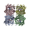


 PDBj
PDBj

 Assembly
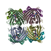

 Mass: 18.015 Da / Num. of mol.: 696 / Source method: isolated from a natural source / Formula: H2O
 Sample preparation
 Processing