 Movie
Movie Controller

[English] 日本語
 Yorodumi
- PDB-2bo8: DISSECTION OF MANNOSYLGLYCERATE SYNTHASE: AN ARCHETYPAL MANNOSYLT... -
+ Open data

- Basic information
| Entry | Database: PDB / ID: 2bo8 | ||||||
|---|---|---|---|---|---|---|---|
| Title | DISSECTION OF MANNOSYLGLYCERATE SYNTHASE: AN ARCHETYPAL MANNOSYLTRANSFERASE | ||||||
 Components Components | MANNOSYLGLYCERATE SYNTHASE | ||||||
 Keywords Keywords | TRANSFERASE / CATALYSIS / GLYCOSYLTRANSFERASE / MANNOSE / STEREOSELECTIVITY | ||||||
| Function / homology |  Function and homology information Function and homology informationmannosylglycerate synthase / mannosylglycerate synthase activity / mannosylglycerate biosynthetic process / hexosyltransferase activity / metal ion binding / identical protein binding Similarity search - Function | ||||||
| Biological species |   RHODOTHERMUS MARINUS (bacteria) RHODOTHERMUS MARINUS (bacteria) | ||||||
| Method |  X-RAY DIFFRACTION / SYNCHROTRON / MOLECULAR REPLACEMENT / Resolution: 2.8 Å X-RAY DIFFRACTION / SYNCHROTRON / MOLECULAR REPLACEMENT / Resolution: 2.8 Å | ||||||
 Authors Authors | Flint, J. / Taylor, E. / Yang, M. / Bolam, D.N. / Tailford, L.E. / Martinez-Fleites, C. / Dodson, E.J. / Davis, B.G. / Gilbert, H.J. / Davies, G.J. | ||||||
 Citation Citation | Journal: Nat. Struct. Mol. Biol. / Year: 2005 Title: Structural dissection and high-throughput screening of mannosylglycerate synthase. Authors: Flint, J. / Taylor, E. / Yang, M. / Bolam, D.N. / Tailford, L.E. / Martinez-Fleites, C. / Dodson, E.J. / Davis, B.G. / Gilbert, H.J. / Davies, G.J. | ||||||
| History |
|
- Structure visualization
| Structure viewer | Molecule: MolmilJmol/JSmol |
|---|
- Downloads & links
-Download
| PDBx/mmCIF format | 2bo8.cif.gz | 767.6 KB | Display | PDBx/mmCIF format |
|---|---|---|---|---|
| PDB format | pdb2bo8.ent.gz | 632.3 KB | Display | PDB format |
| PDBx/mmJSON format | 2bo8.json.gz | Tree view | PDBx/mmJSON format | |
| Others |  Other downloads Other downloads |
-Validation report
| Arichive directory | https://data.pdbj.org/pub/pdb/validation_reports/bo/2bo8ftp://data.pdbj.org/pub/pdb/validation_reports/bo/2bo8 | HTTPS FTP |
|---|
-Related structure data
| Related structure data | 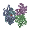 2bo4C  2bo6SC 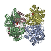 2bo7C C: citing same article ( S: Starting model for refinement |
|---|---|
| Similar structure data |


-Links
 PDBj
PDBj

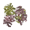
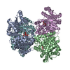
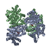
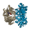
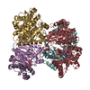
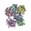
- Assembly
| Deposited unit | 
| ||||||||||||||||||||||||||||||||||||||||||||||||||||||||||||||||||
|---|---|---|---|---|---|---|---|---|---|---|---|---|---|---|---|---|---|---|---|---|---|---|---|---|---|---|---|---|---|---|---|---|---|---|---|---|---|---|---|---|---|---|---|---|---|---|---|---|---|---|---|---|---|---|---|---|---|---|---|---|---|---|---|---|---|---|---|
| 1 | 
| ||||||||||||||||||||||||||||||||||||||||||||||||||||||||||||||||||
| 2 | 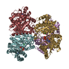
| ||||||||||||||||||||||||||||||||||||||||||||||||||||||||||||||||||
| 3 | 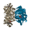
| ||||||||||||||||||||||||||||||||||||||||||||||||||||||||||||||||||
| Unit cell |
| ||||||||||||||||||||||||||||||||||||||||||||||||||||||||||||||||||
| Noncrystallographic symmetry (NCS) | NCS domain:
NCS domain segments: Component-ID: 1 / Ens-ID: 1 / Beg auth comp-ID: SER / Beg label comp-ID: SER / End auth comp-ID: MN / End label comp-ID: MN / Refine code: 1 / Auth seq-ID: 2 - 500 / Label seq-ID: 2
|
-Components
| #1: Protein | Mass: 46186.473 Da / Num. of mol.: 10 Source method: isolated from a genetically manipulated source Source: (gene. exp.) RHODOTHERMUS MARINUS (bacteria) / Plasmid: PET22B / Production host: References: UniProt: Q9RFR0, Transferases; Glycosyltransferases; Hexosyltransferases #2: Chemical | ChemComp-GDX /   Mass: 619.325 Da / Num. of mol.: 10 / Source method: obtained synthetically / Formula: C16H23N5O17P2 Mass: 619.325 Da / Num. of mol.: 10 / Source method: obtained synthetically / Formula: C16H23N5O17P2#3: Chemical | ChemComp-MN /   Mass: 54.938 Da / Num. of mol.: 10 / Source method: obtained synthetically / Formula: Mn Mass: 54.938 Da / Num. of mol.: 10 / Source method: obtained synthetically / Formula: Mn#4: Chemical | ChemComp-CL /   Mass: 35.453 Da / Num. of mol.: 10 / Source method: obtained synthetically / Formula: Cl Mass: 35.453 Da / Num. of mol.: 10 / Source method: obtained synthetically / Formula: Cl#5: Water | ChemComp-HOH / |  Mass: 18.015 Da / Num. of mol.: 308 / Source method: isolated from a natural source / Formula: H2O Mass: 18.015 Da / Num. of mol.: 308 / Source method: isolated from a natural source / Formula: H2O |
|---|
-Experimental details
-Experiment
| Experiment | Method: X-RAY DIFFRACTION |
|---|
- Sample preparation
| Crystal | Density Matthews: 4.2 Å3/Da / Density % sol: 70.7 % |
|---|---|
| Crystal grow | Details: 10 MM NACL, 0.1 M SODIUM ACETATE TRIHYDRATE, PH 4.6, 15% (V/V) MPD |
-Data collection
| Diffraction | Mean temperature: 100 K |
|---|---|
| Diffraction source | Source: SYNCHROTRON / Site: ESRF  / Beamline: ID29 / Wavelength: 0.9791 / Beamline: ID29 / Wavelength: 0.9791 |
| Detector | Type: ADSC CCD / Detector: CCD |
| Radiation | Protocol: SINGLE WAVELENGTH / Monochromatic (M) / Laue (L): M / Scattering type: x-ray |
| Radiation wavelength | Wavelength: 0.9791 Å / Relative weight: 1 |
| Reflection | Resolution: 2.8→40 Å / Num. obs: 175257 / % possible obs: 98 % / Observed criterion σ(I): 2 / Redundancy: 5.3 % / Rmerge(I) obs: 0.1 / Net I/σ(I): 14.5 |
| Reflection shell | Resolution: 2.8→2.95 Å / Redundancy: 4.8 % / Rmerge(I) obs: 0.48 / Mean I/σ(I) obs: 4 / % possible all: 99 |
- Processing
| Software |
| ||||||||||||||||||||||||||||||||||||||||||||||||||||||||||||||||||||||||||||||||||||||||||||||||||||||||||||||||||||||||||||||||||||||||||||||||||||||||||||||||||||||||||||||||||||||
|---|---|---|---|---|---|---|---|---|---|---|---|---|---|---|---|---|---|---|---|---|---|---|---|---|---|---|---|---|---|---|---|---|---|---|---|---|---|---|---|---|---|---|---|---|---|---|---|---|---|---|---|---|---|---|---|---|---|---|---|---|---|---|---|---|---|---|---|---|---|---|---|---|---|---|---|---|---|---|---|---|---|---|---|---|---|---|---|---|---|---|---|---|---|---|---|---|---|---|---|---|---|---|---|---|---|---|---|---|---|---|---|---|---|---|---|---|---|---|---|---|---|---|---|---|---|---|---|---|---|---|---|---|---|---|---|---|---|---|---|---|---|---|---|---|---|---|---|---|---|---|---|---|---|---|---|---|---|---|---|---|---|---|---|---|---|---|---|---|---|---|---|---|---|---|---|---|---|---|---|---|---|---|---|
| Refinement | Method to determine structure: MOLECULAR REPLACEMENT Starting model: PDB ENTRY 2BO6 Resolution: 2.8→74.12 Å / Cor.coef. Fo:Fc: 0.936 / Cor.coef. Fo:Fc free: 0.918 / SU B: 23.342 / SU ML: 0.218 / Cross valid method: THROUGHOUT / ESU R: 0.543 / ESU R Free: 0.276 / Stereochemistry target values: MAXIMUM LIKELIHOOD / Details: HYDROGENS HAVE BEEN ADDED IN THE RIDING POSITIONS.
| ||||||||||||||||||||||||||||||||||||||||||||||||||||||||||||||||||||||||||||||||||||||||||||||||||||||||||||||||||||||||||||||||||||||||||||||||||||||||||||||||||||||||||||||||||||||
| Solvent computation | Ion probe radii: 0.8 Å / Shrinkage radii: 0.8 Å / VDW probe radii: 1.2 Å / Solvent model: MASK | ||||||||||||||||||||||||||||||||||||||||||||||||||||||||||||||||||||||||||||||||||||||||||||||||||||||||||||||||||||||||||||||||||||||||||||||||||||||||||||||||||||||||||||||||||||||
| Displacement parameters | Biso mean: 58.27 Å2
| ||||||||||||||||||||||||||||||||||||||||||||||||||||||||||||||||||||||||||||||||||||||||||||||||||||||||||||||||||||||||||||||||||||||||||||||||||||||||||||||||||||||||||||||||||||||
| Refinement step | Cycle: LAST / Resolution: 2.8→74.12 Å
| ||||||||||||||||||||||||||||||||||||||||||||||||||||||||||||||||||||||||||||||||||||||||||||||||||||||||||||||||||||||||||||||||||||||||||||||||||||||||||||||||||||||||||||||||||||||
| Refine LS restraints |
|