 Movie
Movie Controller

[English] 日本語
 Yorodumi
- PDB-2bo3: Crystal Structure of HP0242, a Hypothetical Protein from Helicoba... -
+ Open data

- Basic information
| Entry | Database: PDB / ID: 2bo3 | ||||||
|---|---|---|---|---|---|---|---|
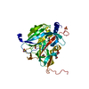
| Title | Crystal Structure of HP0242, a Hypothetical Protein from Helicobacter pylori | ||||||
 Components Components | HYPOTHETICAL PROTEIN HP0242 | ||||||
 Keywords Keywords | STRUCTURAL GENOMICS / UNKNOWN FUNCTION / HELICOBACTER PYLORI / HYPOTHETICAL PROTEIN | ||||||
| Function / homology | HP0242-like domain / HP0242-like fold / Protein of unknown function DUF2018 / HP0242-like superfamily / Domain of unknown function (DUF2018) / Orthogonal Bundle / Mainly Alpha / metal ion binding / DUF2018 family protein Function and homology information Function and homology information | ||||||
| Biological species |   HELICOBACTER PYLORI (bacteria) HELICOBACTER PYLORI (bacteria) | ||||||
| Method |  X-RAY DIFFRACTION / SYNCHROTRON / MAD / Resolution: 2.27 Å X-RAY DIFFRACTION / SYNCHROTRON / MAD / Resolution: 2.27 Å | ||||||
 Authors Authors | Sun, Y.-J. / Tsai, J.-Y. / Chen, B.-T. | ||||||
 Citation Citation | Journal: Proteins: Struct., Funct., Bioinf. / Year: 2006 Title: Crystal Structure of Hp0242, a Hypothetical Protein from Helicobacter Pylori with a Novel Fold Authors: Tsai, J.-Y. / Chen H, B.-T. / Cheng, C. / Chen, H.Y. / Hsiao, N.W. / Liu, P.C. / Sun, Y.-J. | ||||||
| History |
|
- Structure visualization
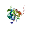
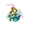
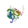
| Structure viewer | Molecule: MolmilJmol/JSmol |
|---|
- Downloads & links
-Download
| PDBx/mmCIF format | 2bo3.cif.gz | 30.9 KB | Display | PDBx/mmCIF format |
|---|---|---|---|---|
| PDB format | pdb2bo3.ent.gz | 20.9 KB | Display | PDB format |
| PDBx/mmJSON format | 2bo3.json.gz | Tree view | PDBx/mmJSON format | |
| Others |  Other downloads Other downloads |
-Validation report
| Arichive directory | https://data.pdbj.org/pub/pdb/validation_reports/bo/2bo3ftp://data.pdbj.org/pub/pdb/validation_reports/bo/2bo3 | HTTPS FTP |
|---|
-Related structure data
| Similar structure data |
|---|

-Links
 PDBj
PDBj- Assembly
| Deposited unit | 
| ||||||||
|---|---|---|---|---|---|---|---|---|---|
| 1 | 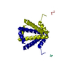
| ||||||||
| Unit cell |
| ||||||||
| Details | REMARK: THE PROTEIN HAS A NOVEL FOLDING AND THE DIMERMIMICS A TIGHT FIGURE-OF-EIGHT KNOT STRUCTURE |
-Components
| #1: Protein | Mass: 11101.640 Da / Num. of mol.: 1 Source method: isolated from a genetically manipulated source Source: (gene. exp.) HELICOBACTER PYLORI (bacteria) / Plasmid: PET30A / Production host: |
|---|---|
| #2: Water | ChemComp-HOH /  Mass: 18.015 Da / Num. of mol.: 44 / Source method: isolated from a natural source / Formula: H2O Mass: 18.015 Da / Num. of mol.: 44 / Source method: isolated from a natural source / Formula: H2O |
-Experimental details
-Experiment
| Experiment | Method: X-RAY DIFFRACTION / Number of used crystals: 1 |
|---|
- Sample preparation
| Crystal | Density Matthews: 2.62 Å3/Da / Density % sol: 53 % |
|---|---|
| Crystal grow | pH: 8.5 / Details: pH 8.50 |
-Data collection
| Diffraction | Mean temperature: 100 K |
|---|---|
| Diffraction source | Source: SYNCHROTRON / Site: SPring-8  / Beamline: BL12B2 / Wavelength: 0.9799 / Beamline: BL12B2 / Wavelength: 0.9799 |
| Detector | Type: ADSC CCD / Detector: CCD / Date: Dec 7, 2004 |
| Radiation | Protocol: MAD / Monochromatic (M) / Laue (L): M / Scattering type: x-ray |
| Radiation wavelength | Wavelength: 0.9799 Å / Relative weight: 1 |
| Reflection | Resolution: 2.27→27.72 Å / Num. obs: 5957 / % possible obs: 93.6 % / Observed criterion σ(I): 2 / Redundancy: 11.9 % / Biso Wilson estimate: 20.7 Å2 / Rmerge(I) obs: 0.05 / Net I/σ(I): 37.2 |
| Reflection shell | Resolution: 2.27→2.35 Å / Redundancy: 11.8 % / Rmerge(I) obs: 0.23 / Mean I/σ(I) obs: 11.9 / % possible all: 92.4 |
- Processing
| Software |
| ||||||||||||||||||||||||||||||||||||||||||||||||||||||||||||||||||||||||||||||||
|---|---|---|---|---|---|---|---|---|---|---|---|---|---|---|---|---|---|---|---|---|---|---|---|---|---|---|---|---|---|---|---|---|---|---|---|---|---|---|---|---|---|---|---|---|---|---|---|---|---|---|---|---|---|---|---|---|---|---|---|---|---|---|---|---|---|---|---|---|---|---|---|---|---|---|---|---|---|---|---|---|---|
| Refinement | Method to determine structure: MAD / Resolution: 2.27→27.72 Å / Rfactor Rfree error: 0.013 / Data cutoff high absF: 311185.28 / Isotropic thermal model: RESTRAINED / Cross valid method: THROUGHOUT / σ(F): 2 / Stereochemistry target values: MAXIMUM LIKELIHOOD
| ||||||||||||||||||||||||||||||||||||||||||||||||||||||||||||||||||||||||||||||||
| Solvent computation | Solvent model: FLAT MODEL / Bsol: 46.9536 Å2 / ksol: 0.369059 e/Å3 | ||||||||||||||||||||||||||||||||||||||||||||||||||||||||||||||||||||||||||||||||
| Displacement parameters | Biso mean: 48.4 Å2
| ||||||||||||||||||||||||||||||||||||||||||||||||||||||||||||||||||||||||||||||||
| Refine analyze |
| ||||||||||||||||||||||||||||||||||||||||||||||||||||||||||||||||||||||||||||||||
| Refinement step | Cycle: LAST / Resolution: 2.27→27.72 Å
| ||||||||||||||||||||||||||||||||||||||||||||||||||||||||||||||||||||||||||||||||
| Refine LS restraints |
| ||||||||||||||||||||||||||||||||||||||||||||||||||||||||||||||||||||||||||||||||
| LS refinement shell | Resolution: 2.2→2.34 Å / Rfactor Rfree error: 0.053 / Total num. of bins used: 6
| ||||||||||||||||||||||||||||||||||||||||||||||||||||||||||||||||||||||||||||||||
| Xplor file |
|
