 Movie
Movie Controller

[English] 日本語
 Yorodumi
- PDB-2bde: Crystal Structure of the cytosolic IMP-GMP specific 5'-nucleotida... -
+ Open data

- Basic information
| Entry | Database: PDB / ID: 2bde | ||||||
|---|---|---|---|---|---|---|---|
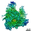
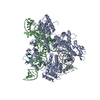
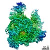
| Title | Crystal Structure of the cytosolic IMP-GMP specific 5'-nucleotidase (lpg0095) from Legionella pneumophila, Northeast Structural Genomics Target LgR1 | ||||||
 Components Components | cytosolic IMP-GMP specific 5'-nucleotidase | ||||||
 Keywords Keywords | DNA BINDING PROTEIN / alpha beta protein / Structural Genomics / PSI / Protein Structure Initiative / Northeast Structural Genomics Consortium / NESG | ||||||
| Function / homology |  Function and homology information Function and homology information5'-nucleotidase activity / identical protein binding / metal ion binding Similarity search - Function | ||||||
| Biological species |   Legionella pneumophila (bacteria) Legionella pneumophila (bacteria) | ||||||
| Method |  X-RAY DIFFRACTION / SYNCHROTRON / MAD / Resolution: 2.9 Å X-RAY DIFFRACTION / SYNCHROTRON / MAD / Resolution: 2.9 Å | ||||||
 Authors Authors | Forouhar, F. / Abashidze, M. / Ho, C.K. / Conover, K. / Acton, T.B. / Montelione, G.T. / Hunt, J.F. / Tong, L. / Northeast Structural Genomics Consortium (NESG) | ||||||
 Citation Citation | Journal: To be Published Title: Crystal Structure of the cytosolic IMP-GMP specific 5'-nucleotidase (lpg0095) from Legionella pneumophila, Northeast Structural Genomics Target LgR1 Authors: Forouhar, F. / Abashidze, M. / Ho, C.K. / Conover, K. / Acton, T.B. / Montelione, G.T. / Hunt, J.F. / Tong, L. | ||||||
| History |
|
- Structure visualization
| Structure viewer | Molecule: MolmilJmol/JSmol |
|---|
- Downloads & links
-Download
| PDBx/mmCIF format | 2bde.cif.gz | 103.3 KB | Display | PDBx/mmCIF format |
|---|---|---|---|---|
| PDB format | pdb2bde.ent.gz | 84.2 KB | Display | PDB format |
| PDBx/mmJSON format | 2bde.json.gz | Tree view | PDBx/mmJSON format | |
| Others |  Other downloads Other downloads |
-Validation report
| Summary document | 2bde_validation.pdf.gz | 441.3 KB | Display | wwPDB validaton report |
|---|---|---|---|---|
| Full document | 2bde_full_validation.pdf.gz | 456.7 KB | Display | |
| Data in XML | 2bde_validation.xml.gz | 20.2 KB | Display | |
| Data in CIF | 2bde_validation.cif.gz | 27.1 KB | Display | |
| Arichive directory | https://data.pdbj.org/pub/pdb/validation_reports/bd/2bdeftp://data.pdbj.org/pub/pdb/validation_reports/bd/2bde | HTTPS FTP |
-Related structure data
| Similar structure data | |
|---|---|
| Other databases |


-Links
 PDBj
PDBj- Assembly
| Deposited unit | 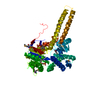
| ||||||||
|---|---|---|---|---|---|---|---|---|---|
| 1 | 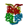
| ||||||||
| 2 | 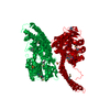
| ||||||||
| 3 | 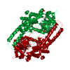
| ||||||||
| Unit cell |
|
-Components
| #1: Protein | Mass: 55158.531 Da / Num. of mol.: 1 Source method: isolated from a genetically manipulated source Source: (gene. exp.) Legionella pneumophila (bacteria) / Strain: Philadelphia 1 / Gene: 3078646 / Plasmid: pET21 / Production host: | ||
|---|---|---|---|
| #2: Chemical |   Mass: 96.063 Da / Num. of mol.: 2 / Source method: obtained synthetically / Formula: SO4 Mass: 96.063 Da / Num. of mol.: 2 / Source method: obtained synthetically / Formula: SO4#3: Water | ChemComp-HOH / |  Mass: 18.015 Da / Num. of mol.: 71 / Source method: isolated from a natural source / Formula: H2O Mass: 18.015 Da / Num. of mol.: 71 / Source method: isolated from a natural source / Formula: H2O |
-Experimental details
-Experiment
| Experiment | Method: X-RAY DIFFRACTION / Number of used crystals: 1 |
|---|
- Sample preparation
| Crystal | Density Matthews: 4.97 Å3/Da / Density % sol: 75.23 % Description: Reflections reported in data collection include friedel pairs |
|---|---|
| Crystal grow | Temperature: 293 K / Method: vapor diffusion, hanging drop / pH: 6.15 Details: 100mM MES, 20% PEG400, 100mM KBr, 5mM DTT, pH 6.15, VAPOR DIFFUSION, HANGING DROP, temperature 293K |
-Data collection
| Diffraction | Mean temperature: 100 K | ||||||||||||
|---|---|---|---|---|---|---|---|---|---|---|---|---|---|
| Diffraction source | Source: SYNCHROTRON / Site: NSLS  / Beamline: X4A / Wavelength: 0.97898, 0.97943, 0.96795 / Beamline: X4A / Wavelength: 0.97898, 0.97943, 0.96795 | ||||||||||||
| Detector | Type: ADSC QUANTUM 4 / Detector: CCD / Date: Sep 16, 2005 / Details: mirrors | ||||||||||||
| Radiation | Monochromator: Si 111 CHANNEL / Protocol: MAD / Monochromatic (M) / Laue (L): M / Scattering type: x-ray | ||||||||||||
| Radiation wavelength |
| ||||||||||||
| Reflection | Resolution: 2.9→30.12 Å / Num. all: 44284 / Num. obs: 44284 / % possible obs: 99.9 % / Observed criterion σ(F): 0 / Observed criterion σ(I): 0 / Redundancy: 24.9 % / Biso Wilson estimate: 70.2 Å2 / Rmerge(I) obs: 0.088 / Rsym value: 0.081 / Net I/σ(I): 29 | ||||||||||||
| Reflection shell | Resolution: 2.9→3 Å / Redundancy: 18.9 % / Rmerge(I) obs: 0.338 / Mean I/σ(I) obs: 10.93 / Num. unique all: 2447 / Rsym value: 0.285 / % possible all: 100 |
- Processing
| Software |
| |||||||||||||||||||||||||
|---|---|---|---|---|---|---|---|---|---|---|---|---|---|---|---|---|---|---|---|---|---|---|---|---|---|---|
| Refinement | Method to determine structure: MAD / Resolution: 2.9→30.12 Å / Rfactor Rfree error: 0.004 / Data cutoff high absF: 475983.63 / Data cutoff low absF: 0 / Isotropic thermal model: OVERALL / Cross valid method: THROUGHOUT / σ(F): 2 / σ(I): 2 / Stereochemistry target values: Engh & Huber / Details: Friedel pairs have been used in refinement
| |||||||||||||||||||||||||
| Solvent computation | Solvent model: FLAT MODEL / Bsol: 20.9387 Å2 / ksol: 0.302233 e/Å3 | |||||||||||||||||||||||||
| Displacement parameters | Biso mean: 49.2 Å2
| |||||||||||||||||||||||||
| Refine analyze |
| |||||||||||||||||||||||||
| Refinement step | Cycle: LAST / Resolution: 2.9→30.12 Å
| |||||||||||||||||||||||||
| Refine LS restraints |
| |||||||||||||||||||||||||
| LS refinement shell | Resolution: 2.9→3.08 Å / Rfactor Rfree error: 0.014 / Total num. of bins used: 6
|
