 Movie
Movie Controller

[English] 日本語
 Yorodumi
- PDB-1y8w: T-To-T(High) quaternary transitions in human hemoglobin: alphaR92... -
+ Open data

- Basic information
| Entry | Database: PDB / ID: 1y8w | ||||||
|---|---|---|---|---|---|---|---|
| Title | T-To-T(High) quaternary transitions in human hemoglobin: alphaR92A oxy (2mM IHP, 20% PEG) (10 test sets) | ||||||
 Components Components |
| ||||||
 Keywords Keywords |  TRANSPORT PROTEIN / HEMOGLOBIN MUTANT / GLOBIN TRANSPORT PROTEIN / HEMOGLOBIN MUTANT / GLOBIN | ||||||
| Function / homology |  Function and homology information Function and homology informationnitric oxide transport / hemoglobin alpha binding / haptoglobin-hemoglobin complex / organic acid binding / cellular oxidant detoxification / hemoglobin binding / renal absorption / hemoglobin complex / oxygen transport / Scavenging of heme from plasma ...nitric oxide transport / hemoglobin alpha binding / haptoglobin-hemoglobin complex / organic acid binding / cellular oxidant detoxification / hemoglobin binding / renal absorption / hemoglobin complex / oxygen transport / Scavenging of heme from plasma / endocytic vesicle lumen / blood vessel diameter maintenance / hydrogen peroxide catabolic process / oxygen carrier activity / Late endosomal microautophagy / Heme signaling / carbon dioxide transport / response to hydrogen peroxide / Erythrocytes take up oxygen and release carbon dioxide / Erythrocytes take up carbon dioxide and release oxygen / Cytoprotection by HMOX1 / platelet aggregation / oxygen binding / regulation of blood pressure / Chaperone Mediated Autophagy / positive regulation of nitric oxide biosynthetic process / tertiary granule lumen / Factors involved in megakaryocyte development and platelet production / blood microparticle / ficolin-1-rich granule lumen / iron ion binding / heme binding / Neutrophil degranulation / extracellular space / extracellular exosome / extracellular region / membrane / metal ion binding / cytosolSimilarity search - Function | ||||||
| Biological species |  Homo sapiens (human) Homo sapiens (human) | ||||||
| Method | X-RAY DIFFRACTION / MOLECULAR REPLACEMENT / Resolution: 2.9 Å | ||||||
 Authors Authors | Kavanaugh, J.S. / Rogers, P.H. / Arnone, A. | ||||||
 Citation Citation | Journal: Biochemistry / Year: 2005 Title: Crystallographic evidence for a new ensemble of ligand-induced allosteric transitions in hemoglobin: the T-to-T(high) quaternary transitions. Authors: Kavanaugh, J.S. / Rogers, P.H. / Arnone, A. | ||||||
| History |
| ||||||
| Remark 600 | HETEROGEN AN IHP MOLECULE WAS NOT ADDED TO THE MODEL SINCE NO SIGNIFICANT ELECTRON DENSITY WAS ...HETEROGEN AN IHP MOLECULE WAS NOT ADDED TO THE MODEL SINCE NO SIGNIFICANT ELECTRON DENSITY WAS FOUND AT ITS BINDING SITE. |
- Structure visualization
| Structure viewer | Molecule: MolmilJmol/JSmol |
|---|
- Downloads & links
-Download
| PDBx/mmCIF format | 1y8w.cif.gz | 128.1 KB | Display | PDBx/mmCIF format |
|---|---|---|---|---|
| PDB format | pdb1y8w.ent.gz | 101 KB | Display | PDB format |
| PDBx/mmJSON format | 1y8w.json.gz | Tree view | PDBx/mmJSON format | |
| Others |  Other downloads Other downloads |
-Validation report
| Arichive directory | https://data.pdbj.org/pub/pdb/validation_reports/y8/1y8wftp://data.pdbj.org/pub/pdb/validation_reports/y8/1y8w | HTTPS FTP |
|---|
-Related structure data
| Related structure data |  1xxtC  1xy0C  1xz5C  1xz7SC  1xzuC  1xzvC  1y09C  1y0aC 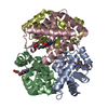 1y0cC 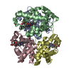 1y0dC 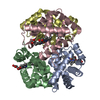 1y0tC 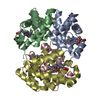 1y0wC  1y22C  1y2zC  1y31C 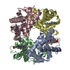 1y35C 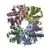 1y45C  1y46C  1y4bC  1y4fC  1y4gC 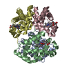 1y4pC 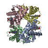 1y4qC  1y4rC  1y4vC  1y5fC  1y5jC  1y5kC  1y7cC  1y7dC  1y7gC  1y7zC  1y83C  1y85C  1ydzC  1ye0C  1ye1C  1ye2C  1yenC  1yeoC  1yeqC  1yeuC  1yevC  1yg5C  1ygdC  1ygfC  1yh9C  1yheC  1yhrC  1yieC  1yihC C: citing same article ( S: Starting model for refinement |
|---|---|
| Similar structure data |


-Links
 PDBj
PDBj


- Assembly
| Deposited unit | 
| ||||||||
|---|---|---|---|---|---|---|---|---|---|
| 1 |
| ||||||||
| Unit cell |
| ||||||||
| Details | the crystallographic asymmetric unit in this entry is an alpha2beta2 tetramer. the crystallographic asymmetric unit and biological unit are equivalent |
-Components
| #1: Protein | Mass: 15096.302 Da / Num. of mol.: 2 / Mutation: V1M, R92A Source method: isolated from a genetically manipulated source Source: (gene. exp.) Homo sapiens (human) / Gene: HBA1 / Production host:  Escherichia coli (E. coli) / References: UniProt: P69905 Escherichia coli (E. coli) / References: UniProt: P69905#2: Protein | Mass: 15890.198 Da / Num. of mol.: 2 / Source method: isolated from a natural source / Source: (natural) Homo sapiens (human) / Tissue: BLOOD / References: UniProt: P68871#3: Chemical | ChemComp-HEM / Heme B  Mass: 616.487 Da / Num. of mol.: 4 / Source method: obtained synthetically / Formula: C34H32FeN4O4 Mass: 616.487 Da / Num. of mol.: 4 / Source method: obtained synthetically / Formula: C34H32FeN4O4#4: Chemical | ChemComp-OXY / Oxygen  Mass: 31.999 Da / Num. of mol.: 4 / Source method: obtained synthetically / Formula: O2 Mass: 31.999 Da / Num. of mol.: 4 / Source method: obtained synthetically / Formula: O2#5: Water | ChemComp-HOH / | Water Mass: 18.015 Da / Num. of mol.: 143 / Source method: isolated from a natural source / Formula: H2O Mass: 18.015 Da / Num. of mol.: 143 / Source method: isolated from a natural source / Formula: H2O |
|---|
-Experimental details
-Experiment
| Experiment | Method: X-RAY DIFFRACTION / Number of used crystals: 1 |
|---|
- Sample preparation
| Crystal | Density Matthews: 2.54 Å3/Da / Density % sol: 51.55 % |
|---|---|
| Crystal grow | Details: 10% PEG 6000, 10 MM POTASSIUM PHOSPHATE, 100 MM POTASSIUM CHLORIDE, 3 MM SODIUM DITHIONITE, 10 MG/ML DEOXYHB, PH 7.0, BATCH, TEMPERATURE 298K, 1 ATM N2. EXPOSING DEOXY CRYSTAL TO LIGAND: A ...Details: 10% PEG 6000, 10 MM POTASSIUM PHOSPHATE, 100 MM POTASSIUM CHLORIDE, 3 MM SODIUM DITHIONITE, 10 MG/ML DEOXYHB, PH 7.0, BATCH, TEMPERATURE 298K, 1 ATM N2. EXPOSING DEOXY CRYSTAL TO LIGAND: A DEOXY CRYSTAL WAS SOAKED (UNDER 1 ATM NITROGEN) IN SUBSTITUTE MOTHER LIQUOR CONTAINING 20% PEG 6000, 10 MM POTASSIUM PHOSPHATE (PH 7.0), 100 MM POTASSIUM CHLORIDE, AND 2.2 MM IHP. THE IHP-STABILIZED CRYSTAL WAS THEN EXPOSED TO 1 ATM OF OXYGEN AT 177K AND DATA WAS COLLECTED AT 169K. |
-Data collection
| Diffraction | Mean temperature: 169 K |
|---|---|
| Diffraction source | Source: ROTATING ANODE / Type: RIGAKU RU200 / Wavelength: 1.5418 / Wavelength: 1.5418 Å |
| Detector | Type: RIGAKU RAXIS IV / Detector: IMAGE PLATE / Date: Sep 20, 2001 / Details: OSMIC MIRRORS |
| Radiation | Monochromator: OSMIC MIRRORS / Protocol: SINGLE WAVELENGTH / Monochromatic (M) / Laue (L): M / Scattering type: x-ray |
| Radiation wavelength | Wavelength: 1.5418 Å / Relative weight: 1 |
| Reflection | Resolution: 2.9→43.9 Å / Num. obs: 13441 / % possible obs: 92.9 % / Observed criterion σ(I): 0 / Redundancy: 2.6 % / Rmerge(I) obs: 0.155 / Net I/σ(I): 3.5 |
| Reflection shell | Resolution: 2.9→3.13 Å / Redundancy: 2.5 % / Rmerge(I) obs: 0.34 / Mean I/σ(I) obs: 2.5 / Num. unique all: 2570 / % possible all: 87.6 |
- Processing
| Software |
| |||||||||||||||||||||||||||||||||||||||||||||||||||||||||||||||||||||||||||||||||||||||||||||||||||||||||
|---|---|---|---|---|---|---|---|---|---|---|---|---|---|---|---|---|---|---|---|---|---|---|---|---|---|---|---|---|---|---|---|---|---|---|---|---|---|---|---|---|---|---|---|---|---|---|---|---|---|---|---|---|---|---|---|---|---|---|---|---|---|---|---|---|---|---|---|---|---|---|---|---|---|---|---|---|---|---|---|---|---|---|---|---|---|---|---|---|---|---|---|---|---|---|---|---|---|---|---|---|---|---|---|---|---|---|
| Refinement | Method to determine structure: MOLECULAR REPLACEMENT Starting model: PDB ENTRY 1XZ7 Resolution: 2.9→10 Å / Cor.coef. Fo:Fc: 0.927 / Cor.coef. Fo:Fc free: 0.837 / SU B: 29.42 / SU ML: 0.559 / Cross valid method: THROUGHOUT / σ(F): 0 / ESU R Free: 0.537 / Stereochemistry target values: MAXIMUM LIKELIHOOD / Details: HYDROGENS HAVE BEEN ADDED IN THE RIDING POSITIONS
| |||||||||||||||||||||||||||||||||||||||||||||||||||||||||||||||||||||||||||||||||||||||||||||||||||||||||
| Solvent computation | Ion probe radii: 0.8 Å / Shrinkage radii: 0.8 Å / VDW probe radii: 1.4 Å / Solvent model: BABINET MODEL WITH MASK | |||||||||||||||||||||||||||||||||||||||||||||||||||||||||||||||||||||||||||||||||||||||||||||||||||||||||
| Displacement parameters | Biso mean: 32.819 Å2
| |||||||||||||||||||||||||||||||||||||||||||||||||||||||||||||||||||||||||||||||||||||||||||||||||||||||||
| Refinement step | Cycle: LAST / Resolution: 2.9→10 Å
| |||||||||||||||||||||||||||||||||||||||||||||||||||||||||||||||||||||||||||||||||||||||||||||||||||||||||
| Refine LS restraints |
| |||||||||||||||||||||||||||||||||||||||||||||||||||||||||||||||||||||||||||||||||||||||||||||||||||||||||
| LS refinement shell | Resolution: 2.9→3.111 Å / Total num. of bins used: 7 /
|
