 Movie
Movie Controller


+ Open data

- Basic information
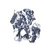
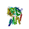
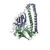
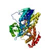
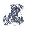
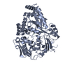
| Entry | Database: PDB / ID: 1wao | ||||||
|---|---|---|---|---|---|---|---|
| Title | PP5 structure | ||||||
 Components Components | SERINE/THREONINE PROTEIN PHOSPHATASE 5 Serine/threonine-specific protein kinase Serine/threonine-specific protein kinase | ||||||
 Keywords Keywords | HYDROLASE / PHOSPHATASE / PROTEIN-PROTEIN INTERACTIONS / TPR / SUPER-HELIX | ||||||
| Function / homology |  Function and homology information Function and homology informationresponse to arachidonic acid / peptidyl-serine dephosphorylation / peptidyl-threonine dephosphorylation / response to morphine / protein folding chaperone complex / myosin phosphatase activity / protein serine/threonine phosphatase activity / protein-serine/threonine phosphatase / phosphatase activity / phosphoprotein phosphatase activity ...response to arachidonic acid / peptidyl-serine dephosphorylation / peptidyl-threonine dephosphorylation / response to morphine / protein folding chaperone complex / myosin phosphatase activity / protein serine/threonine phosphatase activity / protein-serine/threonine phosphatase / phosphatase activity / phosphoprotein phosphatase activity / ESR-mediated signaling / protein dephosphorylation / ADP binding / response to lead ion / Hsp90 protein binding / tau protein binding / Negative regulation of MAPK pathway / double-strand break repair / MAPK cascade / mitotic cell cycle / Recruitment and ATM-mediated phosphorylation of repair and signaling proteins at DNA double strand breaks / positive regulation of canonical NF-kappaB signal transduction / intracellular membrane-bounded organelle / DNA-templated transcription / lipid binding / protein-containing complex / RNA binding / nucleoplasm / ATP binding / identical protein binding / metal ion binding / nucleus / plasma membrane / cytosolSimilarity search - Function | ||||||
| Biological species |  HOMO SAPIENS (human) HOMO SAPIENS (human) | ||||||
| Method | X-RAY DIFFRACTION / SYNCHROTRON / MOLECULAR REPLACEMENT / Resolution: 2.9 Å | ||||||
 Authors Authors | Barford, D. | ||||||
 Citation Citation | Journal: Embo J. / Year: 2005 Title: Molecular Basis for Tpr Domain-Mediated Regulation of Protein Phosphatase 5 Authors: Yang, J. / Roe, S.M. / Cliff, M.J. / Williams, M.A. / Ladbury, J.E. / Cohen, P.T. / Barford, D. | ||||||
| History |
| ||||||
| Remark 700 | SHEET THE SHEET STRUCTURE OF THIS MOLECULE IS BIFURCATED. IN ORDER TO REPRESENT THIS FEATURE IN ... SHEET THE SHEET STRUCTURE OF THIS MOLECULE IS BIFURCATED. IN ORDER TO REPRESENT THIS FEATURE IN THE SHEET RECORDS BELOW, TWO SHEETS ARE DEFINED. |
- Structure visualization
| Structure viewer | Molecule: MolmilJmol/JSmol |
|---|
- Downloads & links
-Download
| PDBx/mmCIF format | 1wao.cif.gz | 369.1 KB | Display | PDBx/mmCIF format |
|---|---|---|---|---|
| PDB format | pdb1wao.ent.gz | 311.7 KB | Display | PDB format |
| PDBx/mmJSON format | 1wao.json.gz | Tree view | PDBx/mmJSON format | |
| Others |  Other downloads Other downloads |
-Validation report
| Arichive directory | https://data.pdbj.org/pub/pdb/validation_reports/wa/1waoftp://data.pdbj.org/pub/pdb/validation_reports/wa/1wao | HTTPS FTP |
|---|
-Related structure data
| Related structure data | |
|---|---|
| Similar structure data |


-Links
 PDBj
PDBj


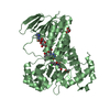
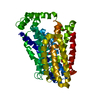
- Assembly
| Deposited unit | 
| ||||||||
|---|---|---|---|---|---|---|---|---|---|
| 1 | 
| ||||||||
| 2 | 
| ||||||||
| 3 | 
| ||||||||
| 4 | 
| ||||||||
| Unit cell |
|
-Components
| #1: Protein | Serine/threonine-specific protein kinase / PP5 / PROTEIN PHOSPHATASE T / PP-T / PPT / PROTEIN PHOSPHATASE 5 Mass: 54566.926 Da / Num. of mol.: 4 Source method: isolated from a genetically manipulated source Source: (gene. exp.) HOMO SAPIENS (human) / Plasmid: PGEX6P / Production host:  ESCHERICHIA COLI (E. coli) / Strain (production host): BL834 ESCHERICHIA COLI (E. coli) / Strain (production host): BL834References: UniProt: P53041, protein-serine/threonine phosphatase #2: Chemical | ChemComp-MN /   Mass: 54.938 Da / Num. of mol.: 8 / Source method: obtained synthetically / Formula: Mn Mass: 54.938 Da / Num. of mol.: 8 / Source method: obtained synthetically / Formula: MnCompound details | CATALYTIC ACTIVITY: PHOSPHOPRO | |
|---|
-Experimental details
-Experiment
| Experiment | Method: X-RAY DIFFRACTION / Number of used crystals: 1 |
|---|
- Sample preparation
| Crystal | Density Matthews: 2.6 Å3/Da / Density % sol: 50 % |
|---|---|
| Crystal grow | Details: PEG 6K 10% |
-Data collection
| Diffraction | Mean temperature: 100 K |
|---|---|
| Diffraction source | Source: SYNCHROTRON / Site: ESRF  / Beamline: ID14-1 / Wavelength: 1 / Beamline: ID14-1 / Wavelength: 1 |
| Detector | Type: ADSC CCD / Detector: CCD / Date: Nov 10, 2002 |
| Radiation | Protocol: SINGLE WAVELENGTH / Monochromatic (M) / Laue (L): M / Scattering type: x-ray |
| Radiation wavelength | Wavelength: 1 Å / Relative weight: 1 |
| Reflection | Resolution: 2.9→50 Å / Num. obs: 53119 / % possible obs: 99.9 % / Observed criterion σ(I): 0 / Redundancy: 6.9 % / Biso Wilson estimate: 102.7 Å2 / Rmerge(I) obs: 0.12 / Net I/σ(I): 3.4 |
| Reflection shell | Resolution: 2.9→3.11 Å / Redundancy: 5.4 % / Rmerge(I) obs: 0.42 / Mean I/σ(I) obs: 1.4 / % possible all: 99.9 |
- Processing
| Software |
| ||||||||||||||||||||||||||||||||||||||||||||||||||||||||||||
|---|---|---|---|---|---|---|---|---|---|---|---|---|---|---|---|---|---|---|---|---|---|---|---|---|---|---|---|---|---|---|---|---|---|---|---|---|---|---|---|---|---|---|---|---|---|---|---|---|---|---|---|---|---|---|---|---|---|---|---|---|---|
| Refinement | Method to determine structure: MOLECULAR REPLACEMENT / Resolution: 2.9→33.78 Å / Rfactor Rfree error: 0.008 / Data cutoff high absF: 1676595.08 / Cross valid method: THROUGHOUT / σ(F): 0
| ||||||||||||||||||||||||||||||||||||||||||||||||||||||||||||
| Solvent computation | Solvent model: CNS BULK SOLVENT MODEL USED / Bsol: 40.0415 Å2 / ksol: 0.270929 e/Å3 | ||||||||||||||||||||||||||||||||||||||||||||||||||||||||||||
| Displacement parameters | Biso mean: 98.43 Å2
| ||||||||||||||||||||||||||||||||||||||||||||||||||||||||||||
| Refine analyze |
| ||||||||||||||||||||||||||||||||||||||||||||||||||||||||||||
| Refinement step | Cycle: LAST / Resolution: 2.9→33.78 Å
| ||||||||||||||||||||||||||||||||||||||||||||||||||||||||||||
| Refine LS restraints |
| ||||||||||||||||||||||||||||||||||||||||||||||||||||||||||||
| LS refinement shell | Highest resolution: 2.9 Å |
