 Movie
Movie Controller


+ Open data

- Basic information
| Entry | Database: PDB / ID: 1vhr | ||||||
|---|---|---|---|---|---|---|---|
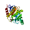
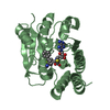
| Title | HUMAN VH1-RELATED DUAL-SPECIFICITY PHOSPHATASE | ||||||
 Components Components | HUMAN VH1-RELATED DUAL-SPECIFICITY PHOSPHATASE VHR | ||||||
 Keywords Keywords |  HYDROLASE / PROTEIN DUAL-SPECIFICITY PHOSPHATASE HYDROLASE / PROTEIN DUAL-SPECIFICITY PHOSPHATASE | ||||||
| Function / homology |  Function and homology informationMAP kinase phosphatase activity / negative regulation of chemotaxis / negative regulation of T cell activation / protein tyrosine/serine/threonine phosphatase activity / positive regulation of focal adhesion disassembly / ERKs are inactivated / negative regulation of JNK cascade / negative regulation of T cell receptor signaling pathway / regulation of focal adhesion assembly / myosin phosphatase activity ...MAP kinase phosphatase activity / negative regulation of chemotaxis / negative regulation of T cell activation / protein tyrosine/serine/threonine phosphatase activity / positive regulation of focal adhesion disassembly / ERKs are inactivated / negative regulation of JNK cascade / negative regulation of T cell receptor signaling pathway / regulation of focal adhesion assembly / myosin phosphatase activity / peptidyl-tyrosine dephosphorylation involved in inactivation of protein kinase activity / negative regulation of MAPK cascade / protein-serine/threonine phosphatase / negative regulation of epidermal growth factor receptor signaling pathway / phosphatase activity / immunological synapse / peptidyl-tyrosine dephosphorylation / dephosphorylation / cellular response to epidermal growth factor stimulus / protein tyrosine kinase binding / cytoskeletal protein binding / positive regulation of mitotic cell cycle / protein-tyrosine-phosphatase / negative regulation of cell migration / protein tyrosine phosphatase activity / negative regulation of ERK1 and ERK2 cascade / receptor tyrosine kinase binding / protein kinase binding / nucleoplasm / nucleus / cytosol / cytoplasm Function and homology informationMAP kinase phosphatase activity / negative regulation of chemotaxis / negative regulation of T cell activation / protein tyrosine/serine/threonine phosphatase activity / positive regulation of focal adhesion disassembly / ERKs are inactivated / negative regulation of JNK cascade / negative regulation of T cell receptor signaling pathway / regulation of focal adhesion assembly / myosin phosphatase activity ...MAP kinase phosphatase activity / negative regulation of chemotaxis / negative regulation of T cell activation / protein tyrosine/serine/threonine phosphatase activity / positive regulation of focal adhesion disassembly / ERKs are inactivated / negative regulation of JNK cascade / negative regulation of T cell receptor signaling pathway / regulation of focal adhesion assembly / myosin phosphatase activity / peptidyl-tyrosine dephosphorylation involved in inactivation of protein kinase activity / negative regulation of MAPK cascade / protein-serine/threonine phosphatase / negative regulation of epidermal growth factor receptor signaling pathway / phosphatase activity / immunological synapse / peptidyl-tyrosine dephosphorylation / dephosphorylation / cellular response to epidermal growth factor stimulus / protein tyrosine kinase binding / cytoskeletal protein binding / positive regulation of mitotic cell cycle / protein-tyrosine-phosphatase / negative regulation of cell migration / protein tyrosine phosphatase activity / negative regulation of ERK1 and ERK2 cascade / receptor tyrosine kinase binding / protein kinase binding / nucleoplasm / nucleus / cytosol / cytoplasmSimilarity search - Function | ||||||
| Biological species |  Homo sapiens (human) Homo sapiens (human) | ||||||
| Method | X-RAY DIFFRACTION / MIR SOFTWARE USED : PHASES STARTING MODEL FOR MOLECULAR REPLACEMENT: NULL / Resolution: 2.1 Å | ||||||
 Authors Authors | Yuvaniyama, J. / Denu, J.M. / Dixon, J.E. / Saper, M.A. | ||||||
 Citation Citation | Journal: Science / Year: 1996 Title: Crystal structure of the dual specificity protein phosphatase VHR. Authors: Yuvaniyama, J. / Denu, J.M. / Dixon, J.E. / Saper, M.A. #1: Journal: J.Biol.Chem. / Year: 1995Title: The Purification and Characterization of a Human Dual-Specific Protein Tyrosine Phosphatase Authors: Denu, J.M. / Zhou, G. / Wu, L. / Zhao, R. / Yuvaniyama, J. / Saper, M.A. / Dixon, J.E. #2: Journal: J.Biol.Chem. / Year: 1994Title: The Catalytic Role of Cys124 in the Dual Specificity Phosphatase Vhr Authors: Zhou, G. / Denu, J.M. / Wu, L. / Dixon, J.E. #3: Journal: Proc.Natl.Acad.Sci.USA / Year: 1992Title: Expression Cloning of a Human Dual-Specificity Phosphatase Authors: Ishibashi, T. / Bottaro, D.P. / Chan, A. / Miki, T. / Aaronson, S.A. | ||||||
| History |
|
- Structure visualization
| Structure viewer | Molecule: MolmilJmol/JSmol |
|---|
- Downloads & links
-Download
| PDBx/mmCIF format | 1vhr.cif.gz | 84 KB | Display | PDBx/mmCIF format |
|---|---|---|---|---|
| PDB format | pdb1vhr.ent.gz | 63.5 KB | Display | PDB format |
| PDBx/mmJSON format | 1vhr.json.gz | Tree view | PDBx/mmJSON format | |
| Others |  Other downloads Other downloads |
-Validation report
| Arichive directory | https://data.pdbj.org/pub/pdb/validation_reports/vh/1vhrftp://data.pdbj.org/pub/pdb/validation_reports/vh/1vhr | HTTPS FTP |
|---|
-Related structure data
| Similar structure data |
|---|


-Links
 PDBj
PDBj


- Assembly
| Deposited unit | 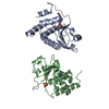
| ||||||||
|---|---|---|---|---|---|---|---|---|---|
| 1 | 
| ||||||||
| 2 | 
| ||||||||
| Unit cell |
| ||||||||
| Noncrystallographic symmetry (NCS) | NCS oper: (Code: given Matrix: (-0.962481, -0.060958, -0.264412), Vector : |
-Components
| #1: Protein | Mass: 20370.078 Da / Num. of mol.: 2 Source method: isolated from a genetically manipulated source Source: (gene. exp.) Homo sapiens (human)Description: ORIGINAL GENE GDB\:DUSP3; VHR, CHROMOSOME MAP POSITION 17Q21 Plasmid: PT7-7-VHR / Species (production host): Escherichia coli / Production host:  Escherichia coli BL21(DE3) (bacteria) / Strain (production host): BL21(DE3) / References: UniProt: P51452, protein-tyrosine-phosphatase Escherichia coli BL21(DE3) (bacteria) / Strain (production host): BL21(DE3) / References: UniProt: P51452, protein-tyrosine-phosphatase#2: Chemical | ChemComp-EPE / | HEPES  Mass: 238.305 Da / Num. of mol.: 1 / Source method: obtained synthetically / Formula: C8H18N2O4S / Comment: pH buffer*YM Mass: 238.305 Da / Num. of mol.: 1 / Source method: obtained synthetically / Formula: C8H18N2O4S / Comment: pH buffer*YM#3: Chemical | ChemComp-SO4 / | Sulfate  Mass: 96.063 Da / Num. of mol.: 1 / Source method: obtained synthetically / Formula: SO4 Mass: 96.063 Da / Num. of mol.: 1 / Source method: obtained synthetically / Formula: SO4#4: Water | ChemComp-HOH / | Water Mass: 18.015 Da / Num. of mol.: 141 / Source method: isolated from a natural source / Formula: H2O Mass: 18.015 Da / Num. of mol.: 141 / Source method: isolated from a natural source / Formula: H2O |
|---|
-Experimental details
-Experiment
| Experiment | Method: X-RAY DIFFRACTION |
|---|
- Sample preparation
| Crystal | Density Matthews: 2.32 Å3/Da / Density % sol: 47.5 % | |||||||||||||||||||||||||||||||||||||||||||||||||
|---|---|---|---|---|---|---|---|---|---|---|---|---|---|---|---|---|---|---|---|---|---|---|---|---|---|---|---|---|---|---|---|---|---|---|---|---|---|---|---|---|---|---|---|---|---|---|---|---|---|---|
| Crystal grow | Temperature: 296 K / Method: vapor diffusion, hanging drop / pH: 7 Details: THE FULL-LENGTH VHR (RESIDUES 2 - 185) WAS CRYSTALLIZED AT A CONCENTRATION OF 5-6 MG/ML WITH 14% POLYETHYLENE GLYCOL 4000, 22.5 MM LITHIUM SULFATE, 50 MM HEPES PH 7.0, AND 0.05% BETA- ...Details: THE FULL-LENGTH VHR (RESIDUES 2 - 185) WAS CRYSTALLIZED AT A CONCENTRATION OF 5-6 MG/ML WITH 14% POLYETHYLENE GLYCOL 4000, 22.5 MM LITHIUM SULFATE, 50 MM HEPES PH 7.0, AND 0.05% BETA-MERCAPTOETHANOL IN A HANGING DROP OF 20 MICROLITERS. THE DROP WAS EQUILIBRATED AGAINST 1 ML OF TWICE THE CONCENTRATION OF PRECIPITANT SOLUTION AT 23 DEGREE CELSIUS., vapor diffusion - hanging drop, temperature 296K | |||||||||||||||||||||||||||||||||||||||||||||||||
| Crystal grow | *PLUS Temperature: 22 ℃ / Method: vapor diffusion, hanging drop | |||||||||||||||||||||||||||||||||||||||||||||||||
| Components of the solutions | *PLUS
|
-Data collection
| Diffraction source | Wavelength: 1.5418 |
|---|---|
| Detector | Type: XUONG-HAMLIN MULTIWIRE / Detector: AREA DETECTOR / Date: Nov 27, 1994 |
| Radiation | Monochromatic (M) / Laue (L): M / Scattering type: x-ray |
| Radiation wavelength | Wavelength: 1.5418 Å / Relative weight: 1 |
| Reflection | Resolution: 2.1→90 Å / Num. obs: 18952 / % possible obs: 86.4 % / Observed criterion σ(I): 0 / Redundancy: 2 % / Rmerge(I) obs: 0.055 |
| Reflection | *PLUS Num. measured all: 41078 |
- Processing
| Software |
| ||||||||||||||||||||||||||||||||||||||||||||||||||||||||||||
|---|---|---|---|---|---|---|---|---|---|---|---|---|---|---|---|---|---|---|---|---|---|---|---|---|---|---|---|---|---|---|---|---|---|---|---|---|---|---|---|---|---|---|---|---|---|---|---|---|---|---|---|---|---|---|---|---|---|---|---|---|---|
| Refinement | Method to determine structure: MIR SOFTWARE USED : PHASES STARTING MODEL FOR MOLECULAR REPLACEMENT: NULL Resolution: 2.1→10 Å / σ(F): 0
| ||||||||||||||||||||||||||||||||||||||||||||||||||||||||||||
| Displacement parameters | Biso mean: 20.7 Å2 | ||||||||||||||||||||||||||||||||||||||||||||||||||||||||||||
| Refinement step | Cycle: LAST / Resolution: 2.1→10 Å
| ||||||||||||||||||||||||||||||||||||||||||||||||||||||||||||
| Refine LS restraints |
| ||||||||||||||||||||||||||||||||||||||||||||||||||||||||||||
| Software | *PLUS Name: X-PLOR / Classification: refinement | ||||||||||||||||||||||||||||||||||||||||||||||||||||||||||||
| Refinement | *PLUS | ||||||||||||||||||||||||||||||||||||||||||||||||||||||||||||
| Solvent computation | *PLUS | ||||||||||||||||||||||||||||||||||||||||||||||||||||||||||||
| Displacement parameters | *PLUS | ||||||||||||||||||||||||||||||||||||||||||||||||||||||||||||
| Refine LS restraints | *PLUS
|
