 Movie
Movie Controller

[English] 日本語
 Yorodumi
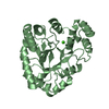
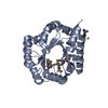
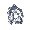
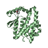
- PDB-1una: UNASSEMBLED VIRUS COAT PROTEIN DIMER, BACTERIOPHAGE RNA-BINDING DIMER -
+ Open data

- Basic information
| Entry | Database: PDB / ID: 1una | ||||||
|---|---|---|---|---|---|---|---|
| Title | UNASSEMBLED VIRUS COAT PROTEIN DIMER, BACTERIOPHAGE RNA-BINDING DIMER | ||||||
 Components Components | GA UNASSEMBLED COAT PROTEIN DIMER | ||||||
 Keywords Keywords | VIRAL PROTEIN / UNASSEMBLED VIRUS COAT PROTEIN DIMER / BACTERIOPHAGE / RNA-BINDING DIMER / TRANSLATIONAL REPRESSOR | ||||||
| Function / homology |  Function and homology information Function and homology informationT=3 icosahedral viral capsid / regulation of translation / structural molecule activity / RNA binding Similarity search - Function | ||||||
| Biological species |  Enterobacteria phage GA (virus) Enterobacteria phage GA (virus) | ||||||
| Method |  X-RAY DIFFRACTION / MOL. REPLACEMENT / Resolution: 2.8 Å X-RAY DIFFRACTION / MOL. REPLACEMENT / Resolution: 2.8 Å | ||||||
 Authors Authors | Ni, C.-Z. / Ely, K.R. | ||||||
 Citation Citation | Journal: Protein Sci. / Year: 1996 Title: Crystal structure of the coat protein from the GA bacteriophage: model of the unassembled dimer. Authors: Ni, C.Z. / White, C.A. / Mitchell, R.S. / Wickersham, J. / Kodandapani, R. / Peabody, D.S. / Ely, K.R. | ||||||
| History |
|
- Structure visualization
| Structure viewer | Molecule: MolmilJmol/JSmol |
|---|
- Downloads & links
-Download
| PDBx/mmCIF format | 1una.cif.gz | 53.6 KB | Display | PDBx/mmCIF format |
|---|---|---|---|---|
| PDB format | pdb1una.ent.gz | 40.6 KB | Display | PDB format |
| PDBx/mmJSON format | 1una.json.gz | Tree view | PDBx/mmJSON format | |
| Others |  Other downloads Other downloads |
-Validation report
| Arichive directory | https://data.pdbj.org/pub/pdb/validation_reports/un/1unaftp://data.pdbj.org/pub/pdb/validation_reports/un/1una | HTTPS FTP |
|---|
-Related structure data
| Related structure data |  1ms2 S: Starting model for refinement |
|---|---|
| Similar structure data |


-Links
 PDBj
PDBj
- Assembly
| Deposited unit | 
| ||||||||
|---|---|---|---|---|---|---|---|---|---|
| 1 |
| ||||||||
| Unit cell |
|
-Components
| #1: Protein | Mass: 13646.364 Da / Num. of mol.: 2 Source method: isolated from a genetically manipulated source Details: THIS BACTERIOPHAGE COAT PROTEIN WAS CRYSTALLIZED AS AN UNASSEMBLED DIMER. IT DID NOT FORM VIRAL CAPSIDS. Source: (gene. exp.) Enterobacteria phage GA (virus) / Genus: Levivirus / Species: Enterobacteria phage BZ13Description: GA COAT SEQUENCE WAS CLONED FROM RNA ISOLATED FROM GA BACTERIOPHAGE PROVIDED BY DR. A. HIRASHIMA, KEIO UNIVERSITY. THE COAT GENE IN THIS DIFFERS AT FOUR SITES FROM THE WIDETYPE PUBLISHED ...Description: GA COAT SEQUENCE WAS CLONED FROM RNA ISOLATED FROM GA BACTERIOPHAGE PROVIDED BY DR. A. HIRASHIMA, KEIO UNIVERSITY. THE COAT GENE IN THIS DIFFERS AT FOUR SITES FROM THE WIDETYPE PUBLISHED SEQUENCE (INOKUCHI ET AL.,(1986) J. BIOCHEM. 99\: 1169) Gene: GA COAT GENE / Plasmid: PUC118 / Gene (production host): GA COAT GENE / Production host:  |
|---|
-Experimental details
-Experiment
| Experiment | Method: X-RAY DIFFRACTION / Number of used crystals: 1 |
|---|
- Sample preparation
| Crystal | Density Matthews: 2.2 Å3/Da / Density % sol: 46 % | ||||||||||||||||||||||||||||||||||||
|---|---|---|---|---|---|---|---|---|---|---|---|---|---|---|---|---|---|---|---|---|---|---|---|---|---|---|---|---|---|---|---|---|---|---|---|---|---|
| Crystal grow | *PLUS Temperature: 13 ℃ / pH: 7.2 / Method: vapor diffusion, hanging drop | ||||||||||||||||||||||||||||||||||||
| Components of the solutions | *PLUS
|
-Data collection
| Diffraction | Mean temperature: 293 K |
|---|---|
| Diffraction source | Source: ROTATING ANODE / Type: RIGAKU RUH2R / Wavelength: 1.5418 |
| Detector | Type: XUONG-HAMLIN MULTIWIRE / Detector: AREA DETECTOR / Date: Dec 3, 1993 |
| Radiation | Monochromator: GRAPHITE(002) / Monochromatic (M) / Laue (L): M / Scattering type: x-ray |
| Radiation wavelength | Wavelength: 1.5418 Å / Relative weight: 1 |
| Reflection | Resolution: 2.8→55 Å / Num. obs: 10793 / % possible obs: 94 % / Redundancy: 3 % / Rmerge(I) obs: 0.066 / Net I/σ(I): 12.9 |
| Reflection shell | Resolution: 2.8→3 Å / Redundancy: 2 % / Rmerge(I) obs: 0.11 / Mean I/σ(I) obs: 5 / % possible all: 81 |
| Reflection | *PLUS Lowest resolution: 4.79 Å / Num. obs: 6248 / Num. measured all: 32352 / Rmerge(I) obs: 0.074 |
| Reflection shell | *PLUS Highest resolution: 2.8 Å / Lowest resolution: 3.02 Å / Num. unique obs: 1157 / Num. measured obs: 3616 / Rmerge(I) obs: 0.123 / Mean I/σ(I) obs: 6.3 |
- Processing
| Software |
| ||||||||||||||||||||||||||||||||||||||||||||||||||||||||||||||||||||||||||||||||||||
|---|---|---|---|---|---|---|---|---|---|---|---|---|---|---|---|---|---|---|---|---|---|---|---|---|---|---|---|---|---|---|---|---|---|---|---|---|---|---|---|---|---|---|---|---|---|---|---|---|---|---|---|---|---|---|---|---|---|---|---|---|---|---|---|---|---|---|---|---|---|---|---|---|---|---|---|---|---|---|---|---|---|---|---|---|---|
| Refinement | Method to determine structure: MOL. REPLACEMENT Starting model: MS2 COAT PROTEIN DIMER (1MS2) 1ms2 Resolution: 2.8→6 Å / σ(F): 2
| ||||||||||||||||||||||||||||||||||||||||||||||||||||||||||||||||||||||||||||||||||||
| Displacement parameters | Biso mean: 17.13 Å2 | ||||||||||||||||||||||||||||||||||||||||||||||||||||||||||||||||||||||||||||||||||||
| Refinement step | Cycle: LAST / Resolution: 2.8→6 Å
| ||||||||||||||||||||||||||||||||||||||||||||||||||||||||||||||||||||||||||||||||||||
| Refine LS restraints |
| ||||||||||||||||||||||||||||||||||||||||||||||||||||||||||||||||||||||||||||||||||||
| Software | *PLUS Name: PROLSQ / Classification: refinement | ||||||||||||||||||||||||||||||||||||||||||||||||||||||||||||||||||||||||||||||||||||
| Refinement | *PLUS | ||||||||||||||||||||||||||||||||||||||||||||||||||||||||||||||||||||||||||||||||||||
| Solvent computation | *PLUS | ||||||||||||||||||||||||||||||||||||||||||||||||||||||||||||||||||||||||||||||||||||
| Displacement parameters | *PLUS |
