 Movie
Movie Controller


+ Open data

- Basic information
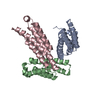
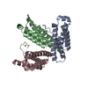
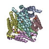
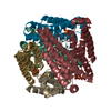
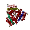
| Entry | Database: PDB / ID: 1t6q | ||||||
|---|---|---|---|---|---|---|---|
| Title | Nickel Superoxide Dismutase (NiSOD) CN-treated Apo Structure | ||||||
 Components Components | Superoxide dismutase [Ni] | ||||||
 Keywords Keywords | OXIDOREDUCTASE / Nickel / 4-helix bundle / hexamer / superoxide dismutase / NiSOD / SOD / apo / cyanide | ||||||
| Function / homology |  Function and homology information Function and homology informationsuperoxide dismutase / superoxide dismutase activity / nickel cation binding / cytoplasm Similarity search - Function | ||||||
| Biological species |  Streptomyces coelicolor (bacteria) Streptomyces coelicolor (bacteria) | ||||||
| Method |  X-RAY DIFFRACTION / SYNCHROTRON / MOLECULAR REPLACEMENT / Resolution: 2.05 Å X-RAY DIFFRACTION / SYNCHROTRON / MOLECULAR REPLACEMENT / Resolution: 2.05 Å | ||||||
 Authors Authors | Barondeau, D.P. / Kassmann, C.J. / Bruns, C.K. / Tainer, J.A. / Getzoff, E.D. | ||||||
 Citation Citation | Journal: Biochemistry / Year: 2004 Title: Nickel superoxide dismutase structure and mechanism. Authors: Barondeau, D.P. / Kassmann, C.J. / Bruns, C.K. / Tainer, J.A. / Getzoff, E.D. | ||||||
| History |
|
- Structure visualization
| Structure viewer | Molecule: MolmilJmol/JSmol |
|---|
- Downloads & links
-Download
| PDBx/mmCIF format | 1t6q.cif.gz | 77.3 KB | Display | PDBx/mmCIF format |
|---|---|---|---|---|
| PDB format | pdb1t6q.ent.gz | 59.2 KB | Display | PDB format |
| PDBx/mmJSON format | 1t6q.json.gz | Tree view | PDBx/mmJSON format | |
| Others |  Other downloads Other downloads |
-Validation report
| Arichive directory | https://data.pdbj.org/pub/pdb/validation_reports/t6/1t6qftp://data.pdbj.org/pub/pdb/validation_reports/t6/1t6q | HTTPS FTP |
|---|
-Related structure data
| Related structure data |  1t6iSC  1t6uC S: Starting model for refinement C: citing same article ( |
|---|---|
| Similar structure data |

-Links
 PDBj
PDBj
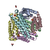
- Assembly
| Deposited unit | 
| ||||||||
|---|---|---|---|---|---|---|---|---|---|
| 1 | 
| ||||||||
| Unit cell |
| ||||||||
| Details | The biological assembly is a hexamer geneated from the trimer in the asymmetric unit by the operation -x,y,-z+1/2 |
-Components
| #1: Protein | Mass: 13241.003 Da / Num. of mol.: 3 / Mutation: L85M Source method: isolated from a genetically manipulated source Source: (gene. exp.) Streptomyces coelicolor (bacteria) / Gene: SODN, SOD1, SCO5254, 2SC7G11.16C / Plasmid: pET26 / Production host: #2: Water | ChemComp-HOH / |  Mass: 18.015 Da / Num. of mol.: 110 / Source method: isolated from a natural source / Formula: H2O Mass: 18.015 Da / Num. of mol.: 110 / Source method: isolated from a natural source / Formula: H2O |
|---|
-Experimental details
-Experiment
| Experiment | Method: X-RAY DIFFRACTION / Number of used crystals: 1 |
|---|
- Sample preparation
| Crystal | Density Matthews: 2.3 Å3/Da / Density % sol: 47 % |
|---|---|
| Crystal grow | Temperature: 295 K / Method: vapor diffusion, hanging drop / pH: 8 Details: MPEG 5000, Hepes, methanol, calcium chloride, pH 8.0, VAPOR DIFFUSION, HANGING DROP, temperature 295K |
-Data collection
| Diffraction | Mean temperature: 100 K |
|---|---|
| Diffraction source | Source: SYNCHROTRON / Site: ALS  / Beamline: 12.3.1 / Wavelength: 1.549784 Å / Beamline: 12.3.1 / Wavelength: 1.549784 Å |
| Detector | Type: ADSC QUANTUM 315 / Detector: CCD / Date: Jan 23, 2004 |
| Radiation | Monochromator: Double Crystal / Protocol: SINGLE WAVELENGTH / Monochromatic (M) / Laue (L): M / Scattering type: x-ray |
| Radiation wavelength | Wavelength: 1.549784 Å / Relative weight: 1 |
| Reflection | Resolution: 2.05→100 Å / Num. all: 23036 / Num. obs: 23036 / % possible obs: 96.4 % / Observed criterion σ(I): -3 / Redundancy: 7.1 % / Biso Wilson estimate: 36.5 Å2 / Rsym value: 0.065 / Net I/σ(I): 24.9 |
| Reflection shell | Resolution: 2.05→2.12 Å / Mean I/σ(I) obs: 5.1 / Num. unique all: 2007 / Rsym value: 0.324 / % possible all: 85.4 |
- Processing
| Software |
| ||||||||||||||||||||||||||||||||||||
|---|---|---|---|---|---|---|---|---|---|---|---|---|---|---|---|---|---|---|---|---|---|---|---|---|---|---|---|---|---|---|---|---|---|---|---|---|---|
| Refinement | Method to determine structure: MOLECULAR REPLACEMENT Starting model: 1T6I Resolution: 2.05→47.83 Å / Rfactor Rfree error: 0.008 / Data cutoff high absF: 401373.55 / Data cutoff high rms absF: 401373.55 / Data cutoff low absF: 0 / Isotropic thermal model: RESTRAINED / Cross valid method: THROUGHOUT / σ(F): 0 / Stereochemistry target values: Engh & Huber
| ||||||||||||||||||||||||||||||||||||
| Solvent computation | Solvent model: FLAT MODEL / Bsol: 49.1048 Å2 / ksol: 0.385898 e/Å3 | ||||||||||||||||||||||||||||||||||||
| Displacement parameters | Biso mean: 48.3 Å2
| ||||||||||||||||||||||||||||||||||||
| Refine analyze |
| ||||||||||||||||||||||||||||||||||||
| Refinement step | Cycle: LAST / Resolution: 2.05→47.83 Å
| ||||||||||||||||||||||||||||||||||||
| Refine LS restraints |
| ||||||||||||||||||||||||||||||||||||
| LS refinement shell | Resolution: 2.05→2.18 Å / Rfactor Rfree error: 0.03 / Total num. of bins used: 6
| ||||||||||||||||||||||||||||||||||||
| Xplor file |
|
