| Entry | Database: PDB / ID: 4ncq
|
|---|
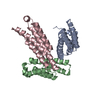
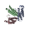
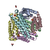
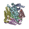
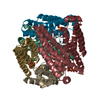
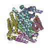
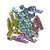
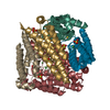
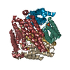
| Title | Crystal structure of NiSOD H1A mutant |
|---|
 Components Components | Superoxide dismutase [Ni] |
|---|
 Keywords Keywords | OXIDOREDUCTASE / antioxidant / hexamer / superoxide dismutase / NiSOD / SOD / metal-binding / Ni-binding / Ni |
|---|
| Function / homology |  Function and homology information Function and homology information
Nickel-containing superoxide dismutase / Superoxide dismutase, Nickel-type / Nickel-containing superoxide dismutase superfamily / Nickel-containing superoxide dismutase / Four Helix Bundle (Hemerythrin (Met), subunit A) / Up-down Bundle / Mainly AlphaSimilarity search - Domain/homology |
|---|
| Biological species |  Streptomyces coelicolor (bacteria) Streptomyces coelicolor (bacteria) |
|---|
| Method |  X-RAY DIFFRACTION / SYNCHROTRON / FOURIER SYNTHESIS / Resolution: 2.08 Å X-RAY DIFFRACTION / SYNCHROTRON / FOURIER SYNTHESIS / Resolution: 2.08 Å |
|---|
 Authors Authors | Guce, A.I. / Garman, S.C. |
|---|
 Citation Citation | Journal: Biochemistry / Year: 2015
Title: Nickel superoxide dismutase: structural and functional roles of His1 and its H-bonding network.
Authors: Ryan, K.C. / Guce, A.I. / Johnson, O.E. / Brunold, T.C. / Cabelli, D.E. / Garman, S.C. / Maroney, M.J. |
|---|
| History | | Deposition | Oct 24, 2013 | Deposition site: RCSB / Processing site: RCSB |
|---|
| Revision 1.0 | Dec 24, 2014 | Provider: repository / Type: Initial release |
|---|
| Revision 1.1 | Apr 8, 2015 | Group: Database references |
|---|
| Revision 1.2 | Nov 15, 2017 | Group: Refinement description / Category: software / Item: _software.name |
|---|
| Revision 1.3 | Sep 20, 2023 | Group: Data collection / Database references / Refinement description
Category: chem_comp_atom / chem_comp_bond ...chem_comp_atom / chem_comp_bond / database_2 / pdbx_initial_refinement_model / struct_ncs_dom_lim / struct_ref_seq_dif
Item: _database_2.pdbx_DOI / _database_2.pdbx_database_accession ..._database_2.pdbx_DOI / _database_2.pdbx_database_accession / _struct_ncs_dom_lim.beg_auth_comp_id / _struct_ncs_dom_lim.beg_label_asym_id / _struct_ncs_dom_lim.beg_label_comp_id / _struct_ncs_dom_lim.beg_label_seq_id / _struct_ncs_dom_lim.end_auth_comp_id / _struct_ncs_dom_lim.end_label_asym_id / _struct_ncs_dom_lim.end_label_comp_id / _struct_ncs_dom_lim.end_label_seq_id / _struct_ref_seq_dif.details |
|---|
|
|---|
 Movie
Movie Controller


 Open data
 Basic information
 Structure visualization
 Downloads & links
 Other downloads


 PDBj
PDBj
 Assembly