- PDB-1s1q: TSG101(UEV) domain in complex with Ubiquitin -
+
データを開く
IDまたはキーワード:
読み込み中...
-
基本情報
登録情報
データベース: PDB / ID: 1s1q
タイトル
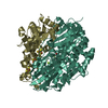
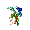
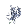
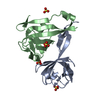
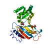
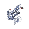
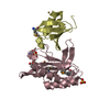
TSG101(UEV) domain in complex with Ubiquitin
要素
Tumor susceptibility gene 101 protein
ubiquitin
キーワード
TRANSLATION / PROTEIN TURNOVER / heterodimer
機能・相同性
機能・相同性情報
positive regulation of viral budding via host ESCRT complex / positive regulation of ubiquitin-dependent endocytosis / extracellular transport / ESCRT I complex / negative regulation of epidermal growth factor-activated receptor activity / regulation of extracellular exosome assembly / viral budding / regulation of MAP kinase activity / : / : ...positive regulation of viral budding via host ESCRT complex / positive regulation of ubiquitin-dependent endocytosis / extracellular transport / ESCRT I complex / negative regulation of epidermal growth factor-activated receptor activity / regulation of extracellular exosome assembly / viral budding / regulation of MAP kinase activity / : / : / protein modification process => GO:0036211 / exosomal secretion / protein transport to vacuole involved in ubiquitin-dependent protein catabolic process via the multivesicular body sorting pathway / positive regulation of exosomal secretion / membrane fission / ubiquitin-dependent protein catabolic process via the multivesicular body sorting pathway / multivesicular body assembly / Flemming body / virion binding / endosome to lysosome transport / viral budding via host ESCRT complex / negative regulation of epidermal growth factor receptor signaling pathway / viral release from host cell / Peptide chain elongation / Selenocysteine synthesis / Formation of a pool of free 40S subunits / autophagosome maturation / Eukaryotic Translation Termination / SRP-dependent cotranslational protein targeting to membrane / Response of EIF2AK4 (GCN2) to amino acid deficiency / Viral mRNA Translation / Nonsense Mediated Decay (NMD) independent of the Exon Junction Complex (EJC) / GTP hydrolysis and joining of the 60S ribosomal subunit / L13a-mediated translational silencing of Ceruloplasmin expression / Major pathway of rRNA processing in the nucleolus and cytosol / keratinocyte differentiation / Nonsense Mediated Decay (NMD) enhanced by the Exon Junction Complex (EJC) / : / multivesicular body / Maturation of protein E / Maturation of protein E / ER Quality Control Compartment (ERQC) / Myoclonic epilepsy of Lafora / FLT3 signaling by CBL mutants / Constitutive Signaling by NOTCH1 HD Domain Mutants / IRAK2 mediated activation of TAK1 complex / Prevention of phagosomal-lysosomal fusion / Alpha-protein kinase 1 signaling pathway / Glycogen synthesis / IRAK1 recruits IKK complex / IRAK1 recruits IKK complex upon TLR7/8 or 9 stimulation / Endosomal Sorting Complex Required For Transport (ESCRT) / Membrane binding and targetting of GAG proteins / Negative regulation of FLT3 / Regulation of TBK1, IKKε (IKBKE)-mediated activation of IRF3, IRF7 / PTK6 Regulates RTKs and Their Effectors AKT1 and DOK1 / Regulation of TBK1, IKKε-mediated activation of IRF3, IRF7 upon TLR3 ligation / IRAK2 mediated activation of TAK1 complex upon TLR7/8 or 9 stimulation / NOTCH2 Activation and Transmission of Signal to the Nucleus / TICAM1,TRAF6-dependent induction of TAK1 complex / TICAM1-dependent activation of IRF3/IRF7 / APC/C:Cdc20 mediated degradation of Cyclin B / Regulation of FZD by ubiquitination / Downregulation of ERBB4 signaling / cytosolic ribosome / APC-Cdc20 mediated degradation of Nek2A / p75NTR recruits signalling complexes / InlA-mediated entry of Listeria monocytogenes into host cells / TRAF6 mediated IRF7 activation in TLR7/8 or 9 signaling / Regulation of pyruvate metabolism / TRAF6-mediated induction of TAK1 complex within TLR4 complex / NF-kB is activated and signals survival / Regulation of innate immune responses to cytosolic DNA / Pexophagy / Downregulation of ERBB2:ERBB3 signaling / NRIF signals cell death from the nucleus / Activated NOTCH1 Transmits Signal to the Nucleus / Regulation of PTEN localization / VLDLR internalisation and degradation / Synthesis of active ubiquitin: roles of E1 and E2 enzymes / ubiquitin binding / Translesion synthesis by REV1 / Regulation of BACH1 activity / TICAM1, RIP1-mediated IKK complex recruitment / MAP3K8 (TPL2)-dependent MAPK1/3 activation / Translesion synthesis by POLK / InlB-mediated entry of Listeria monocytogenes into host cell / Degradation of CDH1 / JNK (c-Jun kinases) phosphorylation and activation mediated by activated human TAK1 / Activation of IRF3, IRF7 mediated by TBK1, IKKε (IKBKE) / Josephin domain DUBs / Downregulation of TGF-beta receptor signaling / Translesion synthesis by POLI / HCMV Late Events / Gap-filling DNA repair synthesis and ligation in GG-NER / Degradation of CRY and PER proteins / IKK complex recruitment mediated by RIP1 / Regulation of activated PAK-2p34 by proteasome mediated degradation / PINK1-PRKN Mediated Mitophagy / TGF-beta receptor signaling in EMT (epithelial to mesenchymal transition) 類似検索 - 分子機能
 ムービー
ムービー コントローラー


 データを開く
 基本情報
基本情報 要素
要素 キーワード
キーワード 機能・相同性情報
 Homo sapiens (ヒト)
 X線回折 /
  データ登録者
 引用
引用 構造の表示
構造の表示 ダウンロードとリンク
 その他のダウンロード


 PDBj
PDBj


 集合体
集合体


 分子量: 63.546 Da / 分子数: 4 / 由来タイプ: 合成 / 式: Cu
 分子量: 96.063 Da / 分子数: 4 / 由来タイプ: 合成 / 式: SO4
 分子量: 60.052 Da / 分子数: 2 / 由来タイプ: 合成 / 式: C2H4O2
 試料調製
試料調製 / ビームライン: 8.3.1 / 波長: 0.97979 Å
 解析
解析