 Movie
Movie Controller

[English] 日本語
 Yorodumi
- PDB-1rer: Crystal structure of the homotrimer of fusion glycoprotein E1 fro... -
+ Open data

- Basic information
| Entry | Database: PDB / ID: 1rer | |||||||||
|---|---|---|---|---|---|---|---|---|---|---|
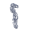
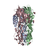
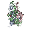
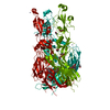
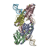
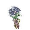
| Title | Crystal structure of the homotrimer of fusion glycoprotein E1 from Semliki Forest Virus. | |||||||||
 Components Components | Structural polyprotein | |||||||||
 Keywords Keywords | VIRAL PROTEIN / Envelope glycoprotein / Membrane fusion / virus. | |||||||||
| Function / homology |  Function and homology information Function and homology informationtogavirin / T=4 icosahedral viral capsid / virion assembly / small molecule binding / host cell endosome / clathrin-dependent endocytosis of virus by host cell / symbiont-mediated suppression of host toll-like receptor signaling pathway / viral translational frameshifting / serine-type endopeptidase activity / fusion of virus membrane with host endosome membrane ...togavirin / T=4 icosahedral viral capsid / virion assembly / small molecule binding / host cell endosome / clathrin-dependent endocytosis of virus by host cell / symbiont-mediated suppression of host toll-like receptor signaling pathway / viral translational frameshifting / serine-type endopeptidase activity / fusion of virus membrane with host endosome membrane / viral envelope / virion attachment to host cell / host cell nucleus / host cell plasma membrane / virion membrane / structural molecule activity / proteolysis / RNA binding / membrane Similarity search - Function | |||||||||
| Biological species |  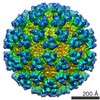 Semliki forest virus Semliki forest virus | |||||||||
| Method |  X-RAY DIFFRACTION / SYNCHROTRON / MAD, MIR / Resolution: 3.2 Å X-RAY DIFFRACTION / SYNCHROTRON / MAD, MIR / Resolution: 3.2 Å | |||||||||
 Authors Authors | Gibbons, D.L. / Vaney, M.C. / Roussel, A. / Vigouroux, A. / Reilly, B. / Kielian, M. / Rey, F.A. | |||||||||
 Citation Citation | Journal: Nature / Year: 2004 Title: Conformational change and protein-protein interactions of the fusion protein of Semliki Forest virus. Authors: Gibbons, D.L. / Vaney, M.C. / Roussel, A. / Vigouroux, A. / Reilly, B. / Lepault, J. / Kielian, M. / Rey, F.A. #1: Journal: Cell(Cambridge,Mass.) / Year: 2001Title: THE FUSION GLYCOPROTEIN SHELL OF SEMLIKI FOREST VIRUS: AN ICOSAHEDRAL ASSEMBLY PRIMED FOR FUSOGENIC ACTIVATION AT ENDOSOMAL PH Authors: LESCAR, J. / ROUSSEL, A. / WIEN, M.W. / NAVAZA, J. / FULLER, S.D. / WENGLER, G. / REY, F.A. #2: Journal: Cell(Cambridge,Mass.) / Year: 2003Title: Visualization of the target-membrane-inserted fusion protein of Semliki Forest virus by combined electron microscopy and crystallography. Authors: Gibbons, D.L. / Erk, I. / Reilly, B. / Navaza, J. / Kielian, M. / Rey, F.A. / Lepault, J. #3: Journal: J.VIROL. / Year: 2002Title: Molecular dissection of the Semliki Forest virus homotrimer reveals two functionally distinct regions of the fusion protein. Authors: Gibbons, D.L. / Lepault, J. | |||||||||
| History |
|
- Structure visualization
| Structure viewer | Molecule: MolmilJmol/JSmol |
|---|
- Downloads & links
-Download
| PDBx/mmCIF format | 1rer.cif.gz | 243 KB | Display | PDBx/mmCIF format |
|---|---|---|---|---|
| PDB format | pdb1rer.ent.gz | 196.6 KB | Display | PDB format |
| PDBx/mmJSON format | 1rer.json.gz | Tree view | PDBx/mmJSON format | |
| Others |  Other downloads Other downloads |
-Validation report
| Arichive directory | https://data.pdbj.org/pub/pdb/validation_reports/re/1rerftp://data.pdbj.org/pub/pdb/validation_reports/re/1rer | HTTPS FTP |
|---|
-Related structure data
| Related structure data | |
|---|---|
| Similar structure data |


-Links
 PDBj
PDBj


- Assembly
| Deposited unit | 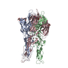
| ||||||||
|---|---|---|---|---|---|---|---|---|---|
| 1 |
| ||||||||
| 2 | 
| ||||||||
| Unit cell |
| ||||||||
| Details | The biological assembly is the homotrimer from the asymmetric unit. |
-Components
-Protein , 1 types, 3 molecules ABC
| #1: Protein | Mass: 42690.125 Da / Num. of mol.: 3 / Fragment: Spike glycoprotein E1 / Source method: isolated from a natural source Details: Contains: Coat protein C (EC 3.4.21.-) (Capsid protein C); Spike glycoprotein E3; Spike glycoprotein E2; Spike glycoprotein E1 Source: (natural) Semliki forest virus / Genus: Alphavirus / References: UniProt: P03315 |
|---|
-Sugars , 3 types, 3 molecules
| #2: Polysaccharide | 2-acetamido-2-deoxy-beta-D-glucopyranose-(1-2)-beta-D-mannopyranose-(1-3)-beta-D-mannopyranose-(1-4) ...2-acetamido-2-deoxy-beta-D-glucopyranose-(1-2)-beta-D-mannopyranose-(1-3)-beta-D-mannopyranose-(1-4)-2-acetamido-2-deoxy-beta-D-glucopyranose-(1-4)-[beta-L-fucopyranose-(1-6)]2-acetamido-2-deoxy-beta-D-glucopyranose Source method: isolated from a genetically manipulated source |
|---|---|
| #3: Polysaccharide | 2-acetamido-2-deoxy-beta-D-glucopyranose-(1-2)-alpha-D-mannopyranose-(1-3)-[alpha-D-mannopyranose- ...2-acetamido-2-deoxy-beta-D-glucopyranose-(1-2)-alpha-D-mannopyranose-(1-3)-[alpha-D-mannopyranose-(1-6)]beta-D-mannopyranose-(1-4)-2-acetamido-2-deoxy-beta-D-glucopyranose-(1-4)-[alpha-L-fucopyranose-(1-6)]2-acetamido-2-deoxy-beta-D-glucopyranose Source method: isolated from a genetically manipulated source |
| #4: Polysaccharide | 2-acetamido-2-deoxy-beta-D-glucopyranose-(1-4)-2-acetamido-2-deoxy-beta-D-glucopyranose Source method: isolated from a genetically manipulated source |
-Non-polymers , 4 types, 147 molecules 


| #5: Chemical |  Mass: 79.904 Da / Num. of mol.: 3 / Source method: obtained synthetically / Formula: Br Mass: 79.904 Da / Num. of mol.: 3 / Source method: obtained synthetically / Formula: Br#6: Chemical | ChemComp-HO /  Mass: 164.930 Da / Num. of mol.: 4 / Source method: obtained synthetically / Formula: Ho Mass: 164.930 Da / Num. of mol.: 4 / Source method: obtained synthetically / Formula: Ho#7: Chemical | ChemComp-PO4 / |  Mass: 94.971 Da / Num. of mol.: 1 / Source method: obtained synthetically / Formula: PO4 Mass: 94.971 Da / Num. of mol.: 1 / Source method: obtained synthetically / Formula: PO4#8: Water | ChemComp-HOH / | Mass: 18.015 Da / Num. of mol.: 139 / Source method: isolated from a natural source / Formula: H2O |
|---|
-Details
| Has protein modification | Y |
|---|
-Experimental details
-Experiment
| Experiment | Method: X-RAY DIFFRACTION / Number of used crystals: 15 |
|---|
- Sample preparation
| Crystal | Density Matthews: 5.15 Å3/Da / Density % sol: 76.1 % | |||||||||||||||||||||||||||||||||||
|---|---|---|---|---|---|---|---|---|---|---|---|---|---|---|---|---|---|---|---|---|---|---|---|---|---|---|---|---|---|---|---|---|---|---|---|---|
| Crystal grow | Temperature: 277 K / Method: vapor diffusion, hanging drop / pH: 4 Details: PEG 400, NaBr, detergent DDAO, HO3+, VAPOR DIFFUSION, HANGING DROP | |||||||||||||||||||||||||||||||||||
| Crystal grow | *PLUS Temperature: 4 ℃ / Method: vapor diffusion, hanging drop | |||||||||||||||||||||||||||||||||||
| Components of the solutions | *PLUS
|
-Data collection
| Diffraction |
| ||||||||||||||||||||
|---|---|---|---|---|---|---|---|---|---|---|---|---|---|---|---|---|---|---|---|---|---|
| Diffraction source |
| ||||||||||||||||||||
| Detector | Type: MARRESEARCH / Detector: CCD / Date: Mar 3, 2003 / Details: Si(111) monochromator | ||||||||||||||||||||
| Radiation | Monochromator: Sagitally focused Si(111) monochromator / Protocol: MAD / Monochromatic (M) / Laue (L): M / Scattering type: x-ray | ||||||||||||||||||||
| Radiation wavelength |
| ||||||||||||||||||||
| Reflection | Resolution: 3.2→20 Å / Num. all: 43822 / Num. obs: 40912 |


- Processing
| Software |
| |||||||||||||||||||||||||
|---|---|---|---|---|---|---|---|---|---|---|---|---|---|---|---|---|---|---|---|---|---|---|---|---|---|---|
| Refinement | Method to determine structure: MAD, MIR / Resolution: 3.2→20 Å / σ(F): 2 / σ(I): 2 / Stereochemistry target values: Engh & Huber
| |||||||||||||||||||||||||
| Refinement step | Cycle: LAST / Resolution: 3.2→20 Å
| |||||||||||||||||||||||||
| LS refinement shell | Resolution: 3.2→3.23 Å
| |||||||||||||||||||||||||
| Refinement | *PLUS % reflection Rfree: 5 % | |||||||||||||||||||||||||
| Solvent computation | *PLUS | |||||||||||||||||||||||||
| Displacement parameters | *PLUS |
