 Movie
Movie Controller


+ Open data

- Basic information
| Entry | Database: PDB / ID: 1rcy | ||||||
|---|---|---|---|---|---|---|---|
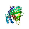
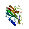
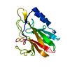
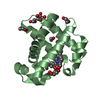
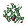
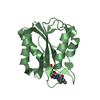
| Title | RUSTICYANIN (RC) FROM THIOBACILLUS FERROOXIDANS | ||||||
 Components Components | RUSTICYANIN | ||||||
 Keywords Keywords | METALLOPROTEIN / COPPER CONTAINING PROTEIN / OXIDATION POTENTIAL / PH STABILITY / REDOX PROTEIN | ||||||
| Function / homology |  Function and homology information Function and homology information | ||||||
| Biological species |  Acidithiobacillus ferrooxidans (bacteria) Acidithiobacillus ferrooxidans (bacteria) | ||||||
| Method |  X-RAY DIFFRACTION / SYNCHROTRON / MULTIWAVELENGTH ANOMALOUS DISPERSION / Resolution: 1.9 Å X-RAY DIFFRACTION / SYNCHROTRON / MULTIWAVELENGTH ANOMALOUS DISPERSION / Resolution: 1.9 Å | ||||||
 Authors Authors | Walter, R.L. / Friedman, A.M. / Ealick, S.E. / Blake II, R.C. / Proctor, P. / Shoham, M. | ||||||
 Citation Citation | Journal: J.Mol.Biol. / Year: 1996 Title: Multiple wavelength anomalous diffraction (MAD) crystal structure of rusticyanin: a highly oxidizing cupredoxin with extreme acid stability. Authors: Walter, R.L. / Ealick, S.E. / Friedman, A.M. / Blake 2nd., R.C. / Proctor, P. / Shoham, M. #1: Journal: To be PublishedTitle: Madprb: A New Suite of Programs for MAD Data Analysis Incorporating Robust Estimation, Maximum Likelihood and Bayesian Inference Authors: Friedman, A.M. / Fischmann, T.O. / Shamoo, Y. / Ealick, S.E. #2: Journal: FEBS Lett. / Year: 1995Title: Complete 13C Assignments for Recombinant Cu(I) Rusticyanin. Prediction of Secondary Structure from Patterns of Chemical Shifts Authors: Toy-Palmer, A. / Prytulla, S. / Dyson, H.J. #3: Journal: Biochemistry / Year: 1995Title: X-Ray Absorption Studies and Homology Modeling Define the Structural Features that Specify the Nature of the Copper Site in Rusticyanin Authors: Grossmann, J.G. / Ingledew, W.J. / Harvey, I. / Strange, R.W. / Hasnain, S.S. #4: Journal: J.Mol.Biol. / Year: 1994Title: Nuclear Magnetic Resonance 15N and 1H Resonance Assignments and Global Fold of Rusticyanin. Insights Into the Ligation and Acid Stability of the Blue Copper Site Authors: Hunt, A.H. / Toy-Palmer, A. / Assa-Munt, N. / Cavanagh, J. / Blake II, R.C. / Dyson, H.J. #5: Journal: J.Mol.Biol. / Year: 1992Title: Crystallization and Preliminary X-Ray Crystallographic Studies of Rusticyanin from Thiobacillus Ferrooxidans Authors: Djebli, A. / Proctor, P. / Blake II, R.C. / Shoham, M. #6: Journal: Geomicrobiol.J. / Year: 1992Title: Respiratory Components in Acidophilic Bacteria that Respire on Iron Authors: Blake II, R.C. / Shute, E.A. / Waskovsky, J. / Harrison Junior, A.P. #7: Journal: Biochemistry / Year: 1991Title: Amino Acid Sequence of the Blue Copper Protein Rusticyanin from Thiobacillus Ferrooxidans Authors: Ronk, M. / Shively, J.E. / Shute, E.A. / Blake II, R.C. #9: Journal: J.Biol.Chem. / Year: 1987Title: Respiratory Enzymes of Thiobacillus Ferrooxidans. A Kinetic Study of Electron Transfer between Iron and Rusticyanin in Sulfate Media Authors: Blake II, R.C. / Shute, E.A. #10: Journal: Biochem.J. / Year: 1978Title: The Purification and Some Properties of Rusticyanin, a Blue Copper Protein Involved in Iron(II) Oxidation from Thiobacillus Ferro-Oxidans Authors: Cox, J.C. / Boxer, D.H. #11: Journal: FEBS Lett. / Year: 1975Title: The Respiratory Chain of Thiobacillus Ferrooxidans: The Reduction of Cytochromes by Fe2+ and the Preliminary Characterization of Rusticyanin, a Novel 'Blue' Copper Protein Authors: Cobley, J.G. / Haddock, B.A. | ||||||
| History |
|
- Structure visualization
| Structure viewer | Molecule: MolmilJmol/JSmol |
|---|
- Downloads & links
-Download
| PDBx/mmCIF format | 1rcy.cif.gz | 45 KB | Display | PDBx/mmCIF format |
|---|---|---|---|---|
| PDB format | pdb1rcy.ent.gz | 30.8 KB | Display | PDB format |
| PDBx/mmJSON format | 1rcy.json.gz | Tree view | PDBx/mmJSON format | |
| Others |  Other downloads Other downloads |
-Validation report
| Arichive directory | https://data.pdbj.org/pub/pdb/validation_reports/rc/1rcyftp://data.pdbj.org/pub/pdb/validation_reports/rc/1rcy | HTTPS FTP |
|---|
-Related structure data
| Similar structure data |
|---|


-Links
 PDBj
PDBj- Assembly
| Deposited unit | 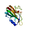
| ||||||||
|---|---|---|---|---|---|---|---|---|---|
| 1 |
| ||||||||
| Unit cell |
|
-Components
| #1: Protein | Mass: 16197.503 Da / Num. of mol.: 1 Source method: isolated from a genetically manipulated source Details: OXIDIZED FORM (CU(II)) Source: (gene. exp.) Acidithiobacillus ferrooxidans (bacteria)Cellular location: PERIPLASM / References: UniProt: P24930, UniProt: P0C918*PLUS |
|---|---|
| #2: Chemical | ChemComp-CU /   Mass: 63.546 Da / Num. of mol.: 1 / Source method: obtained synthetically / Formula: Cu Mass: 63.546 Da / Num. of mol.: 1 / Source method: obtained synthetically / Formula: Cu |
| #3: Water | ChemComp-HOH /  Mass: 18.015 Da / Num. of mol.: 128 / Source method: isolated from a natural source / Formula: H2O Mass: 18.015 Da / Num. of mol.: 128 / Source method: isolated from a natural source / Formula: H2O |
-Experimental details
-Experiment
| Experiment | Method: X-RAY DIFFRACTION / Number of used crystals: 4 |
|---|
- Sample preparation
| Crystal | Density Matthews: 2.16 Å3/Da / Density % sol: 43 % | ||||||||||||||||||||||||||||||||||||||||
|---|---|---|---|---|---|---|---|---|---|---|---|---|---|---|---|---|---|---|---|---|---|---|---|---|---|---|---|---|---|---|---|---|---|---|---|---|---|---|---|---|---|
| Crystal grow | pH: 4.6 / Details: pH 4.6 | ||||||||||||||||||||||||||||||||||||||||
| Crystal grow | *PLUS Temperature: 4 ℃ / Method: vapor diffusion, hanging drop / Details: Djebli, A., (1992) J.Mol.Biol., 227, 581. | ||||||||||||||||||||||||||||||||||||||||
| Components of the solutions | *PLUS
|
-Data collection
| Diffraction | Mean temperature: 277 K |
|---|---|
| Diffraction source | Source: SYNCHROTRON / Site: CHESS  / Beamline: F2 / Wavelength: 1.5418 / Beamline: F2 / Wavelength: 1.5418 |
| Detector | Type: XUONG-HAMLIN MULTIWIRE / Detector: AREA DETECTOR / Date: Oct 20, 1993 / Details: COLLIMATOR |
| Radiation | Monochromator: SI(111) / Monochromatic (M) / Laue (L): M / Scattering type: x-ray |
| Radiation wavelength | Wavelength: 1.5418 Å / Relative weight: 1 |
| Reflection | Highest resolution: 1.9 Å / Num. obs: 9167 / % possible obs: 82 % / Observed criterion σ(I): 2 / Redundancy: 1.5 % / Rmerge(I) obs: 0.02 / Net I/σ(I): 23.5 |
| Reflection shell | Resolution: 1.9→2.05 Å / Redundancy: 1.1 % / Rmerge(I) obs: 0.0383 / Mean I/σ(I) obs: 10.1 / % possible all: 63 |
| Reflection | *PLUS Num. measured all: 14468 |
- Processing
| Software |
| ||||||||||||||||||||||||||||||||||||||||||||||||||||||||||||
|---|---|---|---|---|---|---|---|---|---|---|---|---|---|---|---|---|---|---|---|---|---|---|---|---|---|---|---|---|---|---|---|---|---|---|---|---|---|---|---|---|---|---|---|---|---|---|---|---|---|---|---|---|---|---|---|---|---|---|---|---|---|
| Refinement | Method to determine structure: MULTIWAVELENGTH ANOMALOUS DISPERSION Resolution: 1.9→10 Å / σ(F): 2
| ||||||||||||||||||||||||||||||||||||||||||||||||||||||||||||
| Displacement parameters | Biso mean: 14.3 Å2 | ||||||||||||||||||||||||||||||||||||||||||||||||||||||||||||
| Refinement step | Cycle: LAST / Resolution: 1.9→10 Å
| ||||||||||||||||||||||||||||||||||||||||||||||||||||||||||||
| Refine LS restraints |
| ||||||||||||||||||||||||||||||||||||||||||||||||||||||||||||
| LS refinement shell | Resolution: 1.9→1.99 Å / % reflection obs: 50 % | ||||||||||||||||||||||||||||||||||||||||||||||||||||||||||||
| Xplor file |
| ||||||||||||||||||||||||||||||||||||||||||||||||||||||||||||
| Software | *PLUS Name: X-PLOR / Version: 3.1 / Classification: refinement | ||||||||||||||||||||||||||||||||||||||||||||||||||||||||||||
| Refine LS restraints | *PLUS
|
