 ムービー
ムービー コントローラー


+ データを開く

- 基本情報
基本情報
| 登録情報 | データベース: PDB / ID: 1r7l | ||||||
|---|---|---|---|---|---|---|---|
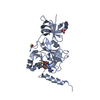
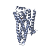
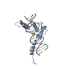
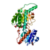
| タイトル | 2.0 A Crystal Structure of a Phage Protein from Bacillus cereus ATCC 14579 | ||||||
 要素 要素 | Phage protein | ||||||
 キーワード キーワード | STRUCTURAL GENOMICS / UNKNOWN FUNCTION / phage protein / two layers alpha-beta sandwich / PSI / Protein Structure Initiative / Midwest Center for Structural Genomics / MCSG | ||||||
| 機能・相同性 | Bacillus phage protein / Bacillus phage protein-like / Bacillus phage protein-like / Phage ABA sandwich domain / Phage ABA sandwich domain / 2-Layer Sandwich / Alpha Beta / Phage protein 機能・相同性情報 機能・相同性情報 | ||||||
| 生物種 |  | ||||||
| 手法 |  X線回折 / シンクロトロン / 多波長異常分散 / 解像度: 2 Å X線回折 / シンクロトロン / 多波長異常分散 / 解像度: 2 Å | ||||||
 データ登録者 データ登録者 | Zhang, R. / Joachimiak, G. / Collart, F. / Joachimiak, A. / Midwest Center for Structural Genomics (MCSG) | ||||||
 引用 引用 | ジャーナル: Proteins / 年: 2006 タイトル: Structure of phage protein BC1872 from Bacillus cereus, a singleton with new fold 著者: Zhang, R. / Joachimiak, G. / Jiang, S. / Cipriani, A. / Collart, F. / Joachimiak, A. | ||||||
| 履歴 |
|
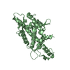
- 構造の表示
構造の表示
| 構造ビューア | 分子: MolmilJmol/JSmol |
|---|
- ダウンロードとリンク
-ダウンロード
| PDBx/mmCIF形式 | 1r7l.cif.gz | 55.5 KB | 表示 | PDBx/mmCIF形式 |
|---|---|---|---|---|
| PDB形式 | pdb1r7l.ent.gz | 40.4 KB | 表示 | PDB形式 |
| PDBx/mmJSON形式 | 1r7l.json.gz | ツリー表示 | PDBx/mmJSON形式 | |
| その他 |  その他のダウンロード その他のダウンロード |
-検証レポート
| アーカイブディレクトリ | https://data.pdbj.org/pub/pdb/validation_reports/r7/1r7lftp://data.pdbj.org/pub/pdb/validation_reports/r7/1r7l | HTTPS FTP |
|---|
-関連構造データ
| 類似構造データ | |
|---|---|
| その他のデータベース |


-リンク
 PDBj
PDBj- 集合体
集合体
| 登録構造単位 | 
| ||||||||
|---|---|---|---|---|---|---|---|---|---|
| 1 |
| ||||||||
| 単位格子 |
| ||||||||
| 詳細 | Molecule A and molecule B represent the dimer in the asymmetric unit. |
-要素
| #1: タンパク質 | 分子量: 12498.479 Da / 分子数: 2 / 由来タイプ: 組換発現 / 由来: (組換発現) #2: 水 | ChemComp-HOH / |  分子量: 18.015 Da / 分子数: 102 / 由来タイプ: 天然 / 式: H2O 分子量: 18.015 Da / 分子数: 102 / 由来タイプ: 天然 / 式: H2O |
|---|
-実験情報
-実験
| 実験 | 手法: X線回折 / 使用した結晶の数: 1 |
|---|
- 試料調製
試料調製
| 結晶 | マシュー密度: 2.64 Å3/Da / 溶媒含有率: 53.36 % |
|---|---|
| 結晶化 | 温度: 298 K / 手法: 蒸気拡散法, シッティングドロップ法 / pH: 6 詳細: 20% PEG 8000, 0.1M MES, 0.2M Ca(OAc)2, pH 6.0, VAPOR DIFFUSION, SITTING DROP, temperature 298K |
-データ収集
| 回折 | 平均測定温度: 100 K | ||||||||||||
|---|---|---|---|---|---|---|---|---|---|---|---|---|---|
| 放射光源 | 由来: シンクロトロン / サイト: APS  / ビームライン: 19-ID / 波長: 0.9795, 0.9798, 0.9465 / ビームライン: 19-ID / 波長: 0.9795, 0.9798, 0.9465 | ||||||||||||
| 検出器 | タイプ: SBC-2 / 検出器: CCD / 日付: 2003年10月20日 / 詳細: mirrors | ||||||||||||
| 放射 | モノクロメーター: Si 111 channel / プロトコル: MAD / 単色(M)・ラウエ(L): M / 散乱光タイプ: x-ray | ||||||||||||
| 放射波長 |
| ||||||||||||
| 反射 | 解像度: 2→50 Å / Num. all: 17716 / Num. obs: 17557 / % possible obs: 99.1 % / Observed criterion σ(F): 4 / Observed criterion σ(I): 4 / 冗長度: 8.12 % / Biso Wilson estimate: 16.2 Å2 / Rmerge(I) obs: 0.098 / Net I/σ(I): 22.3 | ||||||||||||
| 反射 シェル | 解像度: 2→2.07 Å / 冗長度: 7.2 % / Rmerge(I) obs: 0.518 / Mean I/σ(I) obs: 2.25 / % possible all: 95.6 |
- 解析
解析
| ソフトウェア |
| |||||||||||||||||||||||||
|---|---|---|---|---|---|---|---|---|---|---|---|---|---|---|---|---|---|---|---|---|---|---|---|---|---|---|
| 精密化 | 構造決定の手法: 多波長異常分散 / 解像度: 2→24.9 Å / Rfactor Rfree error: 0.007 / Data cutoff high absF: 444377.86 / Data cutoff low absF: 0 / Isotropic thermal model: RESTRAINED / 交差検証法: THROUGHOUT / σ(F): 0 / 立体化学のターゲット値: Engh & Huber / 詳細: Freidel pairs were used in the refinement.
| |||||||||||||||||||||||||
| 溶媒の処理 | 溶媒モデル: FLAT MODEL / Bsol: 60.0238 Å2 / ksol: 0.378568 e/Å3 | |||||||||||||||||||||||||
| 原子変位パラメータ | Biso mean: 33.6 Å2
| |||||||||||||||||||||||||
| Refine analyze |
| |||||||||||||||||||||||||
| 精密化ステップ | サイクル: LAST / 解像度: 2→24.9 Å
| |||||||||||||||||||||||||
| 拘束条件 |
| |||||||||||||||||||||||||
| LS精密化 シェル | 解像度: 2→2.13 Å / Rfactor Rfree error: 0.023 / Total num. of bins used: 6
| |||||||||||||||||||||||||
| Xplor file |
|
