 Movie
Movie Controller

[English] 日本語
 Yorodumi
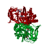
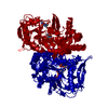
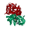
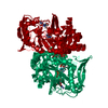
- PDB-1qf5: DESIGN, SYNTHESIS, AND X-RAY CRYSTAL STRUCTURE OF AN ENZYME BOUND... -
+ Open data

- Basic information
| Entry | Database: PDB / ID: 1qf5 | ||||||
|---|---|---|---|---|---|---|---|
| Title | DESIGN, SYNTHESIS, AND X-RAY CRYSTAL STRUCTURE OF AN ENZYME BOUND BISUBSTRATE HYBRID INHIBITOR OF ADENYLOSUCCINATE SYNTHETASE | ||||||
 Components Components | PROTEIN (ADENYLOSUCCINATE SYNTHETASE) | ||||||
 Keywords Keywords | LIGASE / PURINE BIOSYNTHESIS / SYNTHETASE / GTP-BINDING / GTP-HYDROLYSING ENZYMES | ||||||
| Function / homology |  Function and homology information Function and homology informationadenylosuccinate synthase / adenylosuccinate synthase activity / adenosine biosynthetic process / IMP metabolic process / 'de novo' AMP biosynthetic process / nucleobase-containing small molecule interconversion / purine nucleotide biosynthetic process / guanosine tetraphosphate binding / DNA damage response / GTP binding ...adenylosuccinate synthase / adenylosuccinate synthase activity / adenosine biosynthetic process / IMP metabolic process / 'de novo' AMP biosynthetic process / nucleobase-containing small molecule interconversion / purine nucleotide biosynthetic process / guanosine tetraphosphate binding / DNA damage response / GTP binding / magnesium ion binding / membrane / cytoplasm / cytosol Similarity search - Function | ||||||
| Biological species |  | ||||||
| Method |  X-RAY DIFFRACTION / SYNCHROTRON / MOLECULAR REPLACEMENT / Resolution: 2 Å X-RAY DIFFRACTION / SYNCHROTRON / MOLECULAR REPLACEMENT / Resolution: 2 Å | ||||||
 Authors Authors | Hanessian, S. / Lu, P.-P. / Sanceau, J.-Y. / Chemla, P. / Prade, L. / Gohda, K. / Cowan-Jacob, S.W. / Fonne-Pfister, R. | ||||||
 Citation Citation | Journal: Angew.Chem.Int.Ed.Engl. / Year: 1999 Title: An enzyme-bound bisubstrate hybrid inhibitor of adenylosuccinate synthetase Authors: Hanessian, S. / Lu, P.P. / Sanceau, J.Y. / Chemla, P. / Gohda, K. / Fonne-Pfister, R. / Prade, L. / Cowan-Jacob, S.W. #1: Journal: Biochemistry / Year: 1996Title: Refined crystal structure of adenylosuccinate synthetase from Escherichia coli complexed with hydantocidin 5'-phosphate, GDP, HPO4(2-), Mg2+, and hadacidin. Authors: Poland, B.W. / Lee, S.F. / Subramanian, M.V. / Siehl, D.L. / Anderson, R.J. / Fromm, H.J. / Honzatko, R.B. #2: Journal: J.Mol.Biol. / Year: 1995Title: Refined crystal structures of unligated adenylosuccinate synthetase from Escherichia coli. Authors: Silva, M.M. / Poland, B.W. / Hoffman, C.R. / Fromm, H.J. / Honzatko, R.B. #3: Journal: J.Biol.Chem. / Year: 1993 Title: Crystal structure of adenylosuccinate synthetase from Escherichia coli. Evidence for convergent evolution of GTP-binding domains. Authors: Poland, B.W. / Silva, M.M. / Serra, M.A. / Cho, Y. / Kim, K.H. / Harris, E.M. / Honzatko, R.B. | ||||||
| History |
|
- Structure visualization
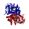
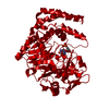
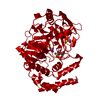
| Structure viewer | Molecule: MolmilJmol/JSmol |
|---|
- Downloads & links
-Download
| PDBx/mmCIF format | 1qf5.cif.gz | 102.1 KB | Display | PDBx/mmCIF format |
|---|---|---|---|---|
| PDB format | pdb1qf5.ent.gz | 75.6 KB | Display | PDB format |
| PDBx/mmJSON format | 1qf5.json.gz | Tree view | PDBx/mmJSON format | |
| Others |  Other downloads Other downloads |
-Validation report
| Arichive directory | https://data.pdbj.org/pub/pdb/validation_reports/qf/1qf5ftp://data.pdbj.org/pub/pdb/validation_reports/qf/1qf5 | HTTPS FTP |
|---|
-Related structure data
| Related structure data |  1qf4C  1juyS C: citing same article ( S: Starting model for refinement |
|---|---|
| Similar structure data |


-Links
 PDBj
PDBj- Assembly
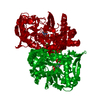
| Deposited unit | 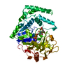
| ||||||||||
|---|---|---|---|---|---|---|---|---|---|---|---|
| 1 | 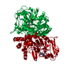
| ||||||||||
| Unit cell |
|
-Components
-Protein , 1 types, 1 molecules A
| #1: Protein | Mass: 47269.598 Da / Num. of mol.: 1 / Source method: isolated from a natural source Details: A GIFT FROM DR. B. BACHURCE 4 (GENETIC CENTER, YALE UNIVERSITY) Source: (natural) |
|---|
-Non-polymers , 5 types, 126 molecules 


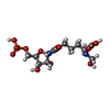

| #2: Chemical | ChemComp-PO4 /  Mass: 94.971 Da / Num. of mol.: 1 / Source method: obtained synthetically / Formula: PO4 Mass: 94.971 Da / Num. of mol.: 1 / Source method: obtained synthetically / Formula: PO4 |
|---|---|
| #3: Chemical | ChemComp-MG /  Mass: 24.305 Da / Num. of mol.: 1 / Source method: obtained synthetically / Formula: Mg Mass: 24.305 Da / Num. of mol.: 1 / Source method: obtained synthetically / Formula: Mg |
| #4: Chemical | ChemComp-GDP /  Type: RNA linking / Mass: 443.201 Da / Num. of mol.: 1 / Source method: obtained synthetically / Formula: C10H15N5O11P2 / Comment: GDP, energy-carrying molecule*YM Type: RNA linking / Mass: 443.201 Da / Num. of mol.: 1 / Source method: obtained synthetically / Formula: C10H15N5O11P2 / Comment: GDP, energy-carrying molecule*YM |
| #5: Chemical | ChemComp-RPL / ( Mass: 459.300 Da / Num. of mol.: 1 / Source method: obtained synthetically / Formula: C13H22N3O13P Mass: 459.300 Da / Num. of mol.: 1 / Source method: obtained synthetically / Formula: C13H22N3O13P |
| #6: Water | ChemComp-HOH / Mass: 18.015 Da / Num. of mol.: 122 / Source method: isolated from a natural source / Formula: H2O |
-Details
| Sequence details | RESIDUE 416 DIVERGES FROM THE DNA SEQUENCE GIVEN IN PIR:A61592 AND IS GLYCINE IN THIS ENTRY. |
|---|
-Experimental details
-Experiment
| Experiment | Method: X-RAY DIFFRACTION / Number of used crystals: 2 |
|---|
- Sample preparation
| Crystal | Density Matthews: 3.24 Å3/Da / Density % sol: 62.01 % | ||||||||||||||||||||
|---|---|---|---|---|---|---|---|---|---|---|---|---|---|---|---|---|---|---|---|---|---|
| Crystal grow | pH: 6.5 / Details: pH 6.5 | ||||||||||||||||||||
| Crystal grow | *PLUS Method: unknown | ||||||||||||||||||||
| Components of the solutions | *PLUS
|
-Data collection
| Diffraction | Mean temperature: 287 K |
|---|---|
| Diffraction source | Source: SYNCHROTRON / Site: ESRF  / Beamline: BM1A / Wavelength: 0.873 / Beamline: BM1A / Wavelength: 0.873 |
| Detector | Type: MARRESEARCH / Detector: IMAGE PLATE / Date: Nov 15, 1996 |
| Radiation | Protocol: SINGLE WAVELENGTH / Monochromatic (M) / Laue (L): M / Scattering type: x-ray |
| Radiation wavelength | Wavelength: 0.873 Å / Relative weight: 1 |
| Reflection | Resolution: 2→8 Å / Num. obs: 31224 / % possible obs: 76.2 % / Redundancy: 4.3 % / Rmerge(I) obs: 0.062 |
- Processing
| Software |
| ||||||||||||||||||||||||||||||||||||||||||||||||||||||||||||
|---|---|---|---|---|---|---|---|---|---|---|---|---|---|---|---|---|---|---|---|---|---|---|---|---|---|---|---|---|---|---|---|---|---|---|---|---|---|---|---|---|---|---|---|---|---|---|---|---|---|---|---|---|---|---|---|---|---|---|---|---|---|
| Refinement | Method to determine structure: MOLECULAR REPLACEMENT Starting model: 1JUY Resolution: 2→6 Å / σ(F): 0
| ||||||||||||||||||||||||||||||||||||||||||||||||||||||||||||
| Displacement parameters | Biso mean: 44 Å2 | ||||||||||||||||||||||||||||||||||||||||||||||||||||||||||||
| Refinement step | Cycle: LAST / Resolution: 2→6 Å
| ||||||||||||||||||||||||||||||||||||||||||||||||||||||||||||
| Refine LS restraints |
|
