 Movie
Movie Controller


+ Open data

- Basic information
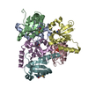
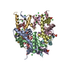
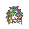
| Entry | Database: PDB / ID: 1q12 | ||||||
|---|---|---|---|---|---|---|---|
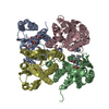
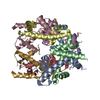
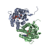
| Title | Crystal Structure of the ATP-bound E. coli MalK | ||||||
 Components Components | Maltose/maltodextrin transport ATP-binding protein malK | ||||||
 Keywords Keywords | TRANSPORT PROTEIN / ATP-binding cassette | ||||||
| Function / homology |  Function and homology information Function and homology informationABC-type maltose transporter / ABC-type maltose transporter activity / negative regulation of maltose transport / enzyme IIA-maltose transporter complex / negative regulation of transmembrane transport / maltose transport complex / maltose transport / maltodextrin transmembrane transport / ATP-binding cassette (ABC) transporter complex, substrate-binding subunit-containing / ATP-binding cassette (ABC) transporter complex ...ABC-type maltose transporter / ABC-type maltose transporter activity / negative regulation of maltose transport / enzyme IIA-maltose transporter complex / negative regulation of transmembrane transport / maltose transport complex / maltose transport / maltodextrin transmembrane transport / ATP-binding cassette (ABC) transporter complex, substrate-binding subunit-containing / ATP-binding cassette (ABC) transporter complex / DNA-binding transcription factor binding / ATP hydrolysis activity / ATP binding / membrane Similarity search - Function | ||||||
| Biological species |  | ||||||
| Method |  X-RAY DIFFRACTION / SYNCHROTRON / MOLECULAR REPLACEMENT / Resolution: 2.6 Å X-RAY DIFFRACTION / SYNCHROTRON / MOLECULAR REPLACEMENT / Resolution: 2.6 Å | ||||||
 Authors Authors | Chen, J. / Lu, G. / Lin, J. / Davidson, A.L. / Quiocho, F.A. | ||||||
 Citation Citation | Journal: Mol.Cell / Year: 2003 Title: A tweezers-like motion of the ATP-binding cassette dimer in an ABC transport cycle Authors: Chen, J. / Lu, G. / Lin, J. / Davidson, A.L. / Quiocho, F.A. | ||||||
| History |
|
- Structure visualization
| Structure viewer | Molecule: MolmilJmol/JSmol |
|---|
- Downloads & links
-Download
| PDBx/mmCIF format | 1q12.cif.gz | 290.1 KB | Display | PDBx/mmCIF format |
|---|---|---|---|---|
| PDB format | pdb1q12.ent.gz | 235.1 KB | Display | PDB format |
| PDBx/mmJSON format | 1q12.json.gz | Tree view | PDBx/mmJSON format | |
| Others |  Other downloads Other downloads |
-Validation report
| Arichive directory | https://data.pdbj.org/pub/pdb/validation_reports/q1/1q12ftp://data.pdbj.org/pub/pdb/validation_reports/q1/1q12 | HTTPS FTP |
|---|
-Related structure data
| Related structure data | 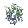 1q1bC 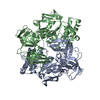 1q1eC 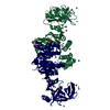 1g29S C: citing same article ( S: Starting model for refinement |
|---|---|
| Similar structure data |


-Links
 PDBj
PDBj

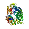
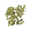
- Assembly
| Deposited unit | 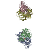
| ||||||||
|---|---|---|---|---|---|---|---|---|---|
| 1 | 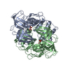
| ||||||||
| 2 | 
| ||||||||
| Unit cell |
|
-Components
| #1: Protein | Mass: 42184.535 Da / Num. of mol.: 4 Source method: isolated from a genetically manipulated source Source: (gene. exp.) #2: Chemical | ChemComp-ATP /   Mass: 507.181 Da / Num. of mol.: 4 / Source method: obtained synthetically / Formula: C10H16N5O13P3 / Comment: ATP, energy-carrying molecule*YM Mass: 507.181 Da / Num. of mol.: 4 / Source method: obtained synthetically / Formula: C10H16N5O13P3 / Comment: ATP, energy-carrying molecule*YM#3: Water | ChemComp-HOH / |  Mass: 18.015 Da / Num. of mol.: 10 / Source method: isolated from a natural source / Formula: H2O Mass: 18.015 Da / Num. of mol.: 10 / Source method: isolated from a natural source / Formula: H2O |
|---|
-Experimental details
-Experiment
| Experiment | Method: X-RAY DIFFRACTION / Number of used crystals: 1 |
|---|
- Sample preparation
| Crystal | Density Matthews: 3.29 Å3/Da / Density % sol: 62.62 % | ||||||||||||||||||||||||||||||||||||||||||||||||||||||||||||
|---|---|---|---|---|---|---|---|---|---|---|---|---|---|---|---|---|---|---|---|---|---|---|---|---|---|---|---|---|---|---|---|---|---|---|---|---|---|---|---|---|---|---|---|---|---|---|---|---|---|---|---|---|---|---|---|---|---|---|---|---|---|
| Crystal grow | Temperature: 273 K / Method: vapor diffusion, hanging drop / pH: 8.8 Details: PEG 4000, glycerol, Sodium Acetate, Tris, pH 8.8, VAPOR DIFFUSION, HANGING DROP, temperature 273K | ||||||||||||||||||||||||||||||||||||||||||||||||||||||||||||
| Crystal grow | *PLUS pH: 8 / Method: vapor diffusion, hanging drop | ||||||||||||||||||||||||||||||||||||||||||||||||||||||||||||
| Components of the solutions | *PLUS
|
-Data collection
| Diffraction | Mean temperature: 200 K |
|---|---|
| Diffraction source | Source: SYNCHROTRON / Site: NSLS  / Beamline: X4A / Wavelength: 0.98 Å / Beamline: X4A / Wavelength: 0.98 Å |
| Detector | Type: ADSC QUANTUM 4 / Detector: CCD / Date: Nov 12, 2001 |
| Radiation | Protocol: SINGLE WAVELENGTH / Monochromatic (M) / Laue (L): M / Scattering type: x-ray |
| Radiation wavelength | Wavelength: 0.98 Å / Relative weight: 1 |
| Reflection | Resolution: 2.6→15 Å / Num. all: 138796 / Num. obs: 124917 / % possible obs: 90 % / Observed criterion σ(F): 1 / Observed criterion σ(I): 1 / Redundancy: 3.9 % / Biso Wilson estimate: 20.5 Å2 / Rmerge(I) obs: 0.1 / Rsym value: 0.106 / Net I/σ(I): 18.5 |
| Reflection shell | Resolution: 2.6→2.69 Å / Redundancy: 2 % / Rmerge(I) obs: 0.283 / Mean I/σ(I) obs: 5.4 / Num. unique all: 8481 / Rsym value: 0.283 / % possible all: 61 |
| Reflection | *PLUS Lowest resolution: 15 Å / % possible obs: 89.9 % / Rmerge(I) obs: 0.106 |
| Reflection shell | *PLUS Highest resolution: 2.6 Å / Lowest resolution: 2.7 Å / % possible obs: 60.1 % / Redundancy: 2.1 % / Num. unique obs: 8481 |
- Processing
| Software |
| ||||||||||||||||||||||||||||||||||||
|---|---|---|---|---|---|---|---|---|---|---|---|---|---|---|---|---|---|---|---|---|---|---|---|---|---|---|---|---|---|---|---|---|---|---|---|---|---|
| Refinement | Method to determine structure: MOLECULAR REPLACEMENT Starting model: 1G29 Resolution: 2.6→14.8 Å / Rfactor Rfree error: 0.003 / Data cutoff high absF: 613412.44 / Data cutoff high rms absF: 613412.44 / Data cutoff low absF: 0 / Isotropic thermal model: RESTRAINED / Cross valid method: THROUGHOUT / σ(F): 0 / σ(I): 0
| ||||||||||||||||||||||||||||||||||||
| Solvent computation | Solvent model: FLAT MODEL / Bsol: 39.6054 Å2 / ksol: 0.331177 e/Å3 | ||||||||||||||||||||||||||||||||||||
| Displacement parameters | Biso mean: 70.1 Å2
| ||||||||||||||||||||||||||||||||||||
| Refine analyze |
| ||||||||||||||||||||||||||||||||||||
| Refinement step | Cycle: LAST / Resolution: 2.6→14.8 Å
| ||||||||||||||||||||||||||||||||||||
| Refine LS restraints |
| ||||||||||||||||||||||||||||||||||||
| LS refinement shell | Resolution: 2.6→2.76 Å / Rfactor Rfree error: 0.013 / Total num. of bins used: 6
| ||||||||||||||||||||||||||||||||||||
| Xplor file |
| ||||||||||||||||||||||||||||||||||||
| Refinement | *PLUS Highest resolution: 2.6 Å / Lowest resolution: 15 Å / % reflection Rfree: 5-8 | ||||||||||||||||||||||||||||||||||||
| Solvent computation | *PLUS | ||||||||||||||||||||||||||||||||||||
| Displacement parameters | *PLUS | ||||||||||||||||||||||||||||||||||||
| Refine LS restraints | *PLUS
|
