 Movie
Movie Controller


+ Open data

- Basic information
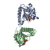
| Entry | Database: PDB / ID: 1pr9 | ||||||
|---|---|---|---|---|---|---|---|
| Title | Human L-Xylulose Reductase Holoenzyme | ||||||
 Components Components | L-XYLULOSE REDUCTASE | ||||||
 Keywords Keywords | OXIDOREDUCTASE / short chain dehydrogenase/reductase / dinucleotide binding domain | ||||||
| Function / homology |  Function and homology information Function and homology informationEssential pentosuria / L-xylulose reductase / L-xylulose reductase (NADPH) activity / Formation of xylulose-5-phosphate / D-glucuronate catabolic process to D-xylulose 5-phosphate / xylulose metabolic process / carbonyl reductase (NADPH) activity / NADP+ metabolic process / D-xylose metabolic process / oxidoreductase activity, acting on NAD(P)H, quinone or similar compound as acceptor ...Essential pentosuria / L-xylulose reductase / L-xylulose reductase (NADPH) activity / Formation of xylulose-5-phosphate / D-glucuronate catabolic process to D-xylulose 5-phosphate / xylulose metabolic process / carbonyl reductase (NADPH) activity / NADP+ metabolic process / D-xylose metabolic process / oxidoreductase activity, acting on NAD(P)H, quinone or similar compound as acceptor / microvillus / brush border / cytoplasmic microtubule / positive regulation of reactive oxygen species metabolic process / glucose metabolic process / mitochondrion / extracellular exosome / identical protein binding / nucleus / plasma membrane / cytosol Similarity search - Function | ||||||
| Biological species |  Homo sapiens (human) Homo sapiens (human) | ||||||
| Method |  X-RAY DIFFRACTION / MOLECULAR REPLACEMENT / Resolution: 1.96 Å X-RAY DIFFRACTION / MOLECULAR REPLACEMENT / Resolution: 1.96 Å | ||||||
 Authors Authors | El-Kabbani, O. / Ishikura, S. / Darmanin, C. / Carbone, V. / Chung, R.P.-T. / Usami, N. / Hara, A. | ||||||
 Citation Citation | Journal: Proteins / Year: 2004 Title: Crystal structure of human L-xylulose reductase holoenzyme: probing the role of Asn107 with site-directed mutagenesis Authors: El-Kabbani, O. / Ishikura, S. / Darmanin, C. / Carbone, V. / Chung, R.P.-T. / Usami, N. / Hara, A. #1: Journal: Acta Crystallogr.,Sect.D / Year: 2002 Title: Crystallization and preliminary crystallographic analysis of human L-xylulose reductase Authors: El-Kabbani, O. / Chung, R.P.-T. / Ishikura, S. / Usami, N. / Nakagawa, J. / Hara, A. | ||||||
| History |
|
- Structure visualization
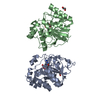
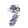
| Structure viewer | Molecule: MolmilJmol/JSmol |
|---|
- Downloads & links
-Download
| PDBx/mmCIF format | 1pr9.cif.gz | 109.4 KB | Display | PDBx/mmCIF format |
|---|---|---|---|---|
| PDB format | pdb1pr9.ent.gz | 84 KB | Display | PDB format |
| PDBx/mmJSON format | 1pr9.json.gz | Tree view | PDBx/mmJSON format | |
| Others |  Other downloads Other downloads |
-Validation report
| Arichive directory | https://data.pdbj.org/pub/pdb/validation_reports/pr/1pr9ftp://data.pdbj.org/pub/pdb/validation_reports/pr/1pr9 | HTTPS FTP |
|---|
-Related structure data
| Similar structure data |
|---|

-Links
 PDBj
PDBj


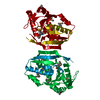
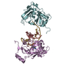
- Assembly
| Deposited unit | 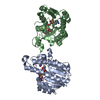
| ||||||||
|---|---|---|---|---|---|---|---|---|---|
| 1 |
| ||||||||
| 2 | 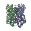
| ||||||||
| Unit cell |
|
-Components
| #1: Protein | Mass: 25941.033 Da / Num. of mol.: 2 Source method: isolated from a genetically manipulated source Source: (gene. exp.) Homo sapiens (human) / Tissue: kidney / Plasmid: pRset / Species (production host): Escherichia coli / Production host:  #2: Chemical |   Mass: 39.098 Da / Num. of mol.: 2 / Source method: obtained synthetically / Formula: K Mass: 39.098 Da / Num. of mol.: 2 / Source method: obtained synthetically / Formula: K#3: Chemical |   Mass: 743.405 Da / Num. of mol.: 2 / Source method: obtained synthetically / Formula: C21H28N7O17P3 Mass: 743.405 Da / Num. of mol.: 2 / Source method: obtained synthetically / Formula: C21H28N7O17P3#4: Chemical |   Mass: 96.987 Da / Num. of mol.: 2 / Source method: obtained synthetically / Formula: H2O4P Mass: 96.987 Da / Num. of mol.: 2 / Source method: obtained synthetically / Formula: H2O4P#5: Water | ChemComp-HOH / |  Mass: 18.015 Da / Num. of mol.: 172 / Source method: isolated from a natural source / Formula: H2O Mass: 18.015 Da / Num. of mol.: 172 / Source method: isolated from a natural source / Formula: H2OHas protein modification | Y | |
|---|
-Experimental details
-Experiment
| Experiment | Method: X-RAY DIFFRACTION / Number of used crystals: 1 |
|---|
- Sample preparation
| Crystal | Density Matthews: 1.9 Å3/Da / Density % sol: 35.2 % / Description: ALL RESIDUES IN THE DIMER WERE DETERMINED | ||||||||||||||||||||||||||||||||||||||||||||||||
|---|---|---|---|---|---|---|---|---|---|---|---|---|---|---|---|---|---|---|---|---|---|---|---|---|---|---|---|---|---|---|---|---|---|---|---|---|---|---|---|---|---|---|---|---|---|---|---|---|---|
| Crystal grow | Temperature: 295 K / Method: vapor diffusion, hanging drop / pH: 6.5 Details: PEG 8000, potassium phosphate, MES, pH 6.5, VAPOR DIFFUSION, HANGING DROP, temperature 295K | ||||||||||||||||||||||||||||||||||||||||||||||||
| Crystal grow | *PLUS Temperature: 295 K / pH: 7.5 / Method: vapor diffusion | ||||||||||||||||||||||||||||||||||||||||||||||||
| Components of the solutions | *PLUS
|
-Data collection
| Diffraction | Mean temperature: 291 K | |||||||||
|---|---|---|---|---|---|---|---|---|---|---|
| Diffraction source | Source: ROTATING ANODE / Type: RIGAKU RU300 / Wavelength: 1.54 / Wavelength: 1.5418 Å | |||||||||
| Detector | Type: MARRESEARCH / Detector: IMAGE PLATE / Date: Dec 1, 2001 / Details: MIRRORS | |||||||||
| Radiation | Monochromator: MAR MIRRORS / Protocol: SINGLE WAVELENGTH / Monochromatic (M) / Laue (L): M / Scattering type: x-ray | |||||||||
| Radiation wavelength |
| |||||||||
| Reflection | Resolution: 1.96→20 Å / Num. all: 34738 / Num. obs: 34738 / % possible obs: 98.7 % / Observed criterion σ(F): 1 / Observed criterion σ(I): 1 / Redundancy: 5 % / Biso Wilson estimate: 22.2 Å2 / Rmerge(I) obs: 0.095 / Net I/σ(I): 18.9 | |||||||||
| Reflection shell | Resolution: 1.98→2.03 Å / Redundancy: 2.5 % / Rmerge(I) obs: 0.275 / Mean I/σ(I) obs: 3.6 / Num. unique all: 3304 / % possible all: 96.61 | |||||||||
| Reflection | *PLUS Num. measured all: 173378 | |||||||||
| Reflection shell | *PLUS Highest resolution: 1.96 Å / % possible obs: 96.61 % / Num. unique obs: 3304 / Mean I/σ(I) obs: 3.5 |
- Processing
| Software |
| |||||||||||||||||||||||||||||||||
|---|---|---|---|---|---|---|---|---|---|---|---|---|---|---|---|---|---|---|---|---|---|---|---|---|---|---|---|---|---|---|---|---|---|---|
| Refinement | Method to determine structure: MOLECULAR REPLACEMENT Starting model: MOUSE LUNG CARBONYL REDUCTASE Resolution: 1.96→10 Å / Num. parameters: 15404 / Num. restraintsaints: 15309 / Isotropic thermal model: isptropic / Cross valid method: THROUGHOUT / σ(F): 0 / σ(I): 0 / Stereochemistry target values: ENGH & HUBER
| |||||||||||||||||||||||||||||||||
| Displacement parameters | Biso mean: 22.2 Å2 | |||||||||||||||||||||||||||||||||
| Refine analyze | Luzzati coordinate error obs: 0.075 Å / Num. disordered residues: 0 / Occupancy sum hydrogen: 0 / Occupancy sum non hydrogen: 3911 | |||||||||||||||||||||||||||||||||
| Refinement step | Cycle: LAST / Resolution: 1.96→10 Å
| |||||||||||||||||||||||||||||||||
| Refine LS restraints |
| |||||||||||||||||||||||||||||||||
| LS refinement shell | Resolution: 1.96→2.03 Å /
| |||||||||||||||||||||||||||||||||
| Software | *PLUS Name: SHELXL / Version: 97 / Classification: refinement | |||||||||||||||||||||||||||||||||
| Refinement | *PLUS Lowest resolution: 10 Å / Rfactor Rwork: 0.1786 | |||||||||||||||||||||||||||||||||
| Solvent computation | *PLUS | |||||||||||||||||||||||||||||||||
| Displacement parameters | *PLUS | |||||||||||||||||||||||||||||||||
| Refine LS restraints | *PLUS
|
