 Movie
Movie Controller

[English] 日本語
 Yorodumi
- PDB-1pnt: CRYSTAL STRUCTURE OF BOVINE HEART PHOSPHOTYROSYL PHOSPHATASE AT 2... -
+ Open data

- Basic information
| Entry | Database: PDB / ID: 1pnt | ||||||
|---|---|---|---|---|---|---|---|
| Title | CRYSTAL STRUCTURE OF BOVINE HEART PHOSPHOTYROSYL PHOSPHATASE AT 2.2 ANGSTROMS RESOLUTION | ||||||
 Components Components | ACID PHOSPHATASE | ||||||
 Keywords Keywords | HYDROLASE | ||||||
| Function / homology |  Function and homology information Function and homology informationacid phosphatase / acid phosphatase activity / non-membrane spanning protein tyrosine phosphatase activity / protein-tyrosine-phosphatase / protein tyrosine phosphatase activity / cytoplasm Similarity search - Function | ||||||
| Biological species |  | ||||||
| Method |  X-RAY DIFFRACTION / Resolution: 2.2 Å X-RAY DIFFRACTION / Resolution: 2.2 Å | ||||||
 Authors Authors | Zhang, M. / Van Etten, R.L. / Stauffacher, C.V. | ||||||
 Citation Citation | Journal: Biochemistry / Year: 1994 Title: Crystal structure of bovine heart phosphotyrosyl phosphatase at 2.2-A resolution. Authors: Zhang, M. / Van Etten, R.L. / Stauffacher, C.V. #1: Journal: J.Mol.Biol. / Year: 1994Title: Crystallization and Preliminary X-Ray Analysis of the Low Molecular Weight Phosphotyrosyl Protein Phosphatase from Bovine Heart Authors: Zhang, M. / Van Etten, R.L. / Lawrence, C.M. / Stauffacher, C.V. | ||||||
| History |
|
- Structure visualization
| Structure viewer | Molecule: MolmilJmol/JSmol |
|---|
- Downloads & links
-Download
| PDBx/mmCIF format | 1pnt.cif.gz | 41.9 KB | Display | PDBx/mmCIF format |
|---|---|---|---|---|
| PDB format | pdb1pnt.ent.gz | 29.4 KB | Display | PDB format |
| PDBx/mmJSON format | 1pnt.json.gz | Tree view | PDBx/mmJSON format | |
| Others |  Other downloads Other downloads |
-Validation report
| Summary document | 1pnt_validation.pdf.gz | 381.3 KB | Display | wwPDB validaton report |
|---|---|---|---|---|
| Full document | 1pnt_full_validation.pdf.gz | 389 KB | Display | |
| Data in XML | 1pnt_validation.xml.gz | 5.8 KB | Display | |
| Data in CIF | 1pnt_validation.cif.gz | 7.7 KB | Display | |
| Arichive directory | https://data.pdbj.org/pub/pdb/validation_reports/pn/1pntftp://data.pdbj.org/pub/pdb/validation_reports/pn/1pnt | HTTPS FTP |
-Related structure data
| Similar structure data |
|---|


-Links
 PDBj
PDBj

- Assembly
| Deposited unit | 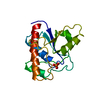
| ||||||||
|---|---|---|---|---|---|---|---|---|---|
| 1 |
| ||||||||
| Unit cell |
| ||||||||
| Atom site foot note | 1: RESIDUES 1 - 3 ARE MODELED IN WEAKER DENSITY AND HAVE CORRESPONDINGLY HIGHER TEMPERATURE FACTORS. |
-Components
| #1: Protein | Mass: 17946.326 Da / Num. of mol.: 1 Source method: isolated from a genetically manipulated source Source: (gene. exp.) |
|---|---|
| #2: Chemical | ChemComp-PO4 /   Mass: 94.971 Da / Num. of mol.: 1 / Source method: obtained synthetically / Formula: PO4 Mass: 94.971 Da / Num. of mol.: 1 / Source method: obtained synthetically / Formula: PO4 |
| Compound details | THE ACTIVE SITE SEQUENCE MOTIF FOR ALL MEMBERS OF THE TYROSINE PHOSPHATASE FAMILY IS CXXXXXRS/T. ...THE ACTIVE SITE SEQUENCE MOTIF FOR ALL MEMBERS OF THE TYROSINE PHOSPHATAS |
-Experimental details
-Experiment
| Experiment | Method: X-RAY DIFFRACTION |
|---|
- Sample preparation
| Crystal | Density Matthews: 2.17 Å3/Da / Density % sol: 43.35 % | ||||||||||||||||||||||||
|---|---|---|---|---|---|---|---|---|---|---|---|---|---|---|---|---|---|---|---|---|---|---|---|---|---|
| Crystal grow | *PLUS pH: 7.5 / Method: vapor diffusion, hanging drop / Details: Zhang, M., (1994) J.Mol.Biol., 238, 281. | ||||||||||||||||||||||||
| Components of the solutions | *PLUS
|
-Data collection
| Radiation | Scattering type: x-ray |
|---|---|
| Radiation wavelength | Relative weight: 1 |
| Reflection | *PLUS Highest resolution: 2.2 Å / % possible obs: 90 % / Redundancy: 3.1 % / Rmerge(I) obs: 0.06 |
- Processing
| Software |
| ||||||||||||||||||||||||||||||||||||||||||||||||||||||||||||
|---|---|---|---|---|---|---|---|---|---|---|---|---|---|---|---|---|---|---|---|---|---|---|---|---|---|---|---|---|---|---|---|---|---|---|---|---|---|---|---|---|---|---|---|---|---|---|---|---|---|---|---|---|---|---|---|---|---|---|---|---|---|
| Refinement | Resolution: 2.2→20 Å / σ(F): 0 Details: RESIDUES 1 - 3 ARE MODELED IN WEAKER DENSITY AND HAVE CORRESPONDINGLY HIGHER TEMPERATURE FACTORS.
| ||||||||||||||||||||||||||||||||||||||||||||||||||||||||||||
| Refinement step | Cycle: LAST / Resolution: 2.2→20 Å
| ||||||||||||||||||||||||||||||||||||||||||||||||||||||||||||
| Refine LS restraints |
| ||||||||||||||||||||||||||||||||||||||||||||||||||||||||||||
| Software | *PLUS Name: TNT / Classification: refinement | ||||||||||||||||||||||||||||||||||||||||||||||||||||||||||||
| Refine LS restraints | *PLUS
|
