 Movie
Movie Controller


+ Open data

- Basic information
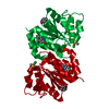
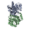
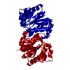
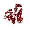
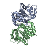
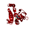
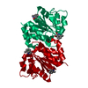
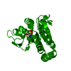
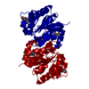
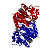
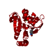
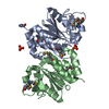
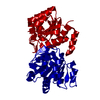
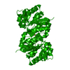
| Entry | Database: PDB / ID: 1pdw | ||||||
|---|---|---|---|---|---|---|---|
| Title | Crystal structure of human DJ-1, P 1 21 1 space group | ||||||
 Components Components | DJ-1 | ||||||
 Keywords Keywords | PROTEIN BINDING / DJ-1 | ||||||
| Function / homology |  Function and homology information Function and homology informationtyrosine 3-monooxygenase activator activity / cellular response to glyoxal / L-dopa decarboxylase activator activity / peptidyl-cysteine deglycation / peptidyl-arginine deglycation / peptidyl-lysine deglycation / protein deglycation, methylglyoxal removal / : / detoxification of hydrogen peroxide / methylglyoxal catabolic process to lactate ...tyrosine 3-monooxygenase activator activity / cellular response to glyoxal / L-dopa decarboxylase activator activity / peptidyl-cysteine deglycation / peptidyl-arginine deglycation / peptidyl-lysine deglycation / protein deglycation, methylglyoxal removal / : / detoxification of hydrogen peroxide / methylglyoxal catabolic process to lactate / guanine deglycation, methylglyoxal removal / cellular detoxification of methylglyoxal / regulation of supramolecular fiber organization / negative regulation of death-inducing signaling complex assembly / negative regulation of TRAIL-activated apoptotic signaling pathway / positive regulation of pyrroline-5-carboxylate reductase activity / positive regulation of tyrosine 3-monooxygenase activity / positive regulation of L-dopa biosynthetic process / positive regulation of L-dopa decarboxylase activity / negative regulation of hydrogen peroxide-induced neuron intrinsic apoptotic signaling pathway / glyoxalase (glycolic acid-forming) activity / negative regulation of protein K48-linked deubiquitination / negative regulation of ubiquitin-specific protease activity / protein deglycation, glyoxal removal / glycolate biosynthetic process / guanine deglycation, glyoxal removal / glyoxal metabolic process / negative regulation of nitrosative stress-induced intrinsic apoptotic signaling pathway / detection of oxidative stress / guanine deglycation / positive regulation of NAD(P)H oxidase activity / detoxification of mercury ion / protein deglycase / methylglyoxal metabolic process / positive regulation of mitochondrial electron transport, NADH to ubiquinone / mercury ion binding / hydrogen peroxide metabolic process / protein deglycase activity / positive regulation of dopamine biosynthetic process / oxidoreductase activity, acting on peroxide as acceptor / superoxide dismutase copper chaperone activity / positive regulation of autophagy of mitochondrion / positive regulation of acute inflammatory response to antigenic stimulus / positive regulation of superoxide dismutase activity / lactate biosynthetic process / negative regulation of cysteine-type endopeptidase activity involved in apoptotic signaling pathway / cellular detoxification of aldehyde / protein deglycosylation / small protein activating enzyme binding / positive regulation of transcription regulatory region DNA binding / Hydrolases; Acting on ester bonds; Thioester hydrolases / peroxiredoxin activity / regulation of oxidative stress-induced neuron intrinsic apoptotic signaling pathway / negative regulation of ubiquitin-protein transferase activity / detoxification of copper ion / negative regulation of protein acetylation / positive regulation of oxidative stress-induced intrinsic apoptotic signaling pathway / negative regulation of oxidative stress-induced neuron intrinsic apoptotic signaling pathway / negative regulation of protein sumoylation / positive regulation of androgen receptor activity / membrane hyperpolarization / negative regulation of protein export from nucleus / regulation of androgen receptor signaling pathway / cupric ion binding / oxygen sensor activity / negative regulation of intrinsic apoptotic signaling pathway in response to hydrogen peroxide / insulin secretion / ubiquitin-like protein conjugating enzyme binding / Hydrolases; Acting on carbon-nitrogen bonds, other than peptide bonds; In linear amides / positive regulation of reactive oxygen species biosynthetic process / nuclear androgen receptor binding / dopamine uptake involved in synaptic transmission / ubiquitin-specific protease binding / cuprous ion binding / cytokine binding / membrane depolarization / single fertilization / negative regulation of endoplasmic reticulum stress-induced intrinsic apoptotic signaling pathway / regulation of neuron apoptotic process / negative regulation of reactive oxygen species biosynthetic process / negative regulation of oxidative stress-induced intrinsic apoptotic signaling pathway / negative regulation of proteasomal ubiquitin-dependent protein catabolic process / negative regulation of protein ubiquitination / activation of protein kinase B activity / adult locomotory behavior / SUMOylation of transcription cofactors / negative regulation of protein phosphorylation / regulation of mitochondrial membrane potential / mitochondrion organization / negative regulation of protein binding / positive regulation of interleukin-8 production / negative regulation of extrinsic apoptotic signaling pathway / adherens junction / kinase binding / negative regulation of protein kinase activity / positive regulation of DNA-binding transcription factor activity / Late endosomal microautophagy / positive regulation of protein-containing complex assembly / PML body / mitochondrial intermembrane space Similarity search - Function | ||||||
| Biological species |  Homo sapiens (human) Homo sapiens (human) | ||||||
| Method |  X-RAY DIFFRACTION / SYNCHROTRON / MOLECULAR REPLACEMENT / Resolution: 2.2 Å X-RAY DIFFRACTION / SYNCHROTRON / MOLECULAR REPLACEMENT / Resolution: 2.2 Å | ||||||
 Authors Authors | Tao, X. / Tong, L. | ||||||
 Citation Citation | Journal: J.Biol.Chem. / Year: 2003 Title: Crystal Structure of Human DJ-1, a Protein Associated with Early Onset Parkinson's Disease. Authors: Tao, X. / Tong, L. | ||||||
| History |
|
- Structure visualization
| Structure viewer | Molecule: MolmilJmol/JSmol |
|---|
- Downloads & links
-Download
| PDBx/mmCIF format | 1pdw.cif.gz | 300.8 KB | Display | PDBx/mmCIF format |
|---|---|---|---|---|
| PDB format | pdb1pdw.ent.gz | 248.7 KB | Display | PDB format |
| PDBx/mmJSON format | 1pdw.json.gz | Tree view | PDBx/mmJSON format | |
| Others |  Other downloads Other downloads |
-Validation report
| Summary document | 1pdw_validation.pdf.gz | 494.9 KB | Display | wwPDB validaton report |
|---|---|---|---|---|
| Full document | 1pdw_full_validation.pdf.gz | 527.1 KB | Display | |
| Data in XML | 1pdw_validation.xml.gz | 65.9 KB | Display | |
| Data in CIF | 1pdw_validation.cif.gz | 91.9 KB | Display | |
| Arichive directory | https://data.pdbj.org/pub/pdb/validation_reports/pd/1pdwftp://data.pdbj.org/pub/pdb/validation_reports/pd/1pdw | HTTPS FTP |
-Related structure data


-Links
 PDBj
PDBj

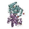
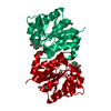
- Assembly
| Deposited unit | 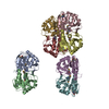
| ||||||||
|---|---|---|---|---|---|---|---|---|---|
| 1 | 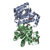
| ||||||||
| 2 | 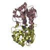
| ||||||||
| 3 | 
| ||||||||
| 4 | 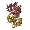
| ||||||||
| Unit cell |
|
-Components
| #1: Protein | Mass: 21175.785 Da / Num. of mol.: 8 Source method: isolated from a genetically manipulated source Source: (gene. exp.) Homo sapiens (human) / Production host:  #2: Water | ChemComp-HOH / |  Mass: 18.015 Da / Num. of mol.: 970 / Source method: isolated from a natural source / Formula: H2O Mass: 18.015 Da / Num. of mol.: 970 / Source method: isolated from a natural source / Formula: H2O |
|---|
-Experimental details
-Experiment
| Experiment | Method: X-RAY DIFFRACTION / Number of used crystals: 1 |
|---|
- Sample preparation
| Crystal | Density Matthews: 1.97 Å3/Da / Density % sol: 37.68 % | ||||||||||||||||||||||||||||||
|---|---|---|---|---|---|---|---|---|---|---|---|---|---|---|---|---|---|---|---|---|---|---|---|---|---|---|---|---|---|---|---|
| Crystal grow | Temperature: 277 K / Method: vapor diffusion, hanging drop / pH: 8 Details: 10% PEG3350, 100 mM Tris, pH 8.0, VAPOR DIFFUSION, HANGING DROP, temperature 277K | ||||||||||||||||||||||||||||||
| Crystal grow | *PLUS Temperature: 4 ℃ / Method: vapor diffusion | ||||||||||||||||||||||||||||||
| Components of the solutions | *PLUS
|
-Data collection
| Diffraction | Mean temperature: 100 K |
|---|---|
| Diffraction source | Source: SYNCHROTRON / Site: NSLS  / Beamline: X4A / Wavelength: 0.9724 Å / Beamline: X4A / Wavelength: 0.9724 Å |
| Detector | Type: ADSC QUANTUM 4 / Detector: CCD / Date: Apr 9, 2003 |
| Radiation | Protocol: SINGLE WAVELENGTH / Monochromatic (M) / Laue (L): M / Scattering type: x-ray |
| Radiation wavelength | Wavelength: 0.9724 Å / Relative weight: 1 |
| Reflection | Resolution: 2.2→20 Å / Num. all: 69228 / Num. obs: 67190 / % possible obs: 99.5 % / Observed criterion σ(F): 0 / Observed criterion σ(I): 0 / Biso Wilson estimate: 7.1 Å2 / Rmerge(I) obs: 0.094 / Rsym value: 0.094 |
| Reflection shell | Resolution: 2.2→2.28 Å / Rmerge(I) obs: 0.231 / % possible all: 99.5 |
| Reflection | *PLUS Num. measured all: 211982 |
- Processing
| Software |
| ||||||||||||||||||||||||||||||||||||
|---|---|---|---|---|---|---|---|---|---|---|---|---|---|---|---|---|---|---|---|---|---|---|---|---|---|---|---|---|---|---|---|---|---|---|---|---|---|
| Refinement | Method to determine structure: MOLECULAR REPLACEMENT / Resolution: 2.2→19.91 Å / Rfactor Rfree error: 0.004 / Isotropic thermal model: RESTRAINED / Cross valid method: THROUGHOUT / σ(F): 2 / Stereochemistry target values: Engh & Huber
| ||||||||||||||||||||||||||||||||||||
| Solvent computation | Solvent model: FLAT MODEL / Bsol: 47.1871 Å2 / ksol: 0.32813 e/Å3 | ||||||||||||||||||||||||||||||||||||
| Displacement parameters | Biso mean: 19.5 Å2
| ||||||||||||||||||||||||||||||||||||
| Refine analyze | Luzzati coordinate error free: 0.31 Å / Luzzati sigma a free: 0.24 Å | ||||||||||||||||||||||||||||||||||||
| Refinement step | Cycle: LAST / Resolution: 2.2→19.91 Å
| ||||||||||||||||||||||||||||||||||||
| Refine LS restraints |
| ||||||||||||||||||||||||||||||||||||
| LS refinement shell | Resolution: 2.2→2.28 Å / Rfactor Rfree error: 0.013 / Total num. of bins used: 10
| ||||||||||||||||||||||||||||||||||||
| Xplor file |
| ||||||||||||||||||||||||||||||||||||
| Refinement | *PLUS Highest resolution: 2.2 Å / Lowest resolution: 20 Å / Rfactor Rwork: 0.18 | ||||||||||||||||||||||||||||||||||||
| Solvent computation | *PLUS | ||||||||||||||||||||||||||||||||||||
| Displacement parameters | *PLUS | ||||||||||||||||||||||||||||||||||||
| Refine LS restraints | *PLUS
|
