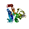
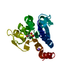
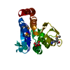
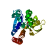
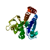
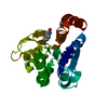
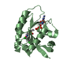
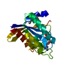
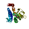
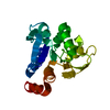
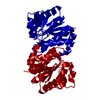
[English] 日本語
Yorodumi
- PDB-1j42: Crystal Structure of Human DJ-1 -

+
Open data


ID or keywords:

Loading...

-
Basic information

Entry
Database: PDB / ID: 1j42
TitleCrystal Structure of Human DJ-1
ComponentsRNA-binding protein regulatory subunit
KeywordsRNA BINDING PROTEIN / Parkinson's disease
Function / homology
Function and homology information


tyrosine 3-monooxygenase activator activity / cellular response to glyoxal / L-dopa decarboxylase activator activity / peptidyl-cysteine deglycation / peptidyl-arginine deglycation / peptidyl-lysine deglycation / protein deglycation, methylglyoxal removal / glutathione deglycation / detoxification of hydrogen peroxide / methylglyoxal catabolic process to lactate ...tyrosine 3-monooxygenase activator activity / cellular response to glyoxal / L-dopa decarboxylase activator activity / peptidyl-cysteine deglycation / peptidyl-arginine deglycation / peptidyl-lysine deglycation / protein deglycation, methylglyoxal removal / glutathione deglycation / detoxification of hydrogen peroxide / methylglyoxal catabolic process to lactate / guanine deglycation, methylglyoxal removal / cellular detoxification of methylglyoxal / regulation of supramolecular fiber organization / negative regulation of death-inducing signaling complex assembly / negative regulation of TRAIL-activated apoptotic signaling pathway / positive regulation of pyrroline-5-carboxylate reductase activity / positive regulation of tyrosine 3-monooxygenase activity / positive regulation of L-dopa biosynthetic process / positive regulation of L-dopa decarboxylase activity / negative regulation of hydrogen peroxide-induced neuron intrinsic apoptotic signaling pathway / glyoxalase (glycolic acid-forming) activity / negative regulation of protein K48-linked deubiquitination / negative regulation of ubiquitin-specific protease activity / protein deglycation, glyoxal removal / glycolate biosynthetic process / guanine deglycation, glyoxal removal / glyoxal metabolic process / negative regulation of nitrosative stress-induced intrinsic apoptotic signaling pathway / detection of oxidative stress / guanine deglycation / detoxification of mercury ion / protein deglycase / methylglyoxal metabolic process / positive regulation of mitochondrial electron transport, NADH to ubiquinone / mercury ion binding / oxidoreductase activity, acting on peroxide as acceptor / protein deglycase activity / positive regulation of acute inflammatory response to antigenic stimulus / positive regulation of dopamine biosynthetic process / superoxide dismutase copper chaperone activity / positive regulation of NAD(P)H oxidase activity / positive regulation of autophagy of mitochondrion / lactate biosynthetic process / negative regulation of cysteine-type endopeptidase activity involved in apoptotic signaling pathway / cellular detoxification of aldehyde / positive regulation of superoxide dismutase activity / small protein activating enzyme binding / Hydrolases; Acting on ester bonds; Thioester hydrolases / regulation of oxidative stress-induced neuron intrinsic apoptotic signaling pathway / negative regulation of ubiquitin-protein transferase activity / peroxiredoxin activity / detoxification of copper ion / negative regulation of protein acetylation / negative regulation of oxidative stress-induced neuron intrinsic apoptotic signaling pathway / positive regulation of transcription regulatory region DNA binding / positive regulation of oxidative stress-induced intrinsic apoptotic signaling pathway / positive regulation of androgen receptor activity / membrane hyperpolarization / protein deglycosylation / negative regulation of protein sumoylation / oxygen sensor activity / regulation of androgen receptor signaling pathway / negative regulation of protein export from nucleus / negative regulation of intrinsic apoptotic signaling pathway in response to hydrogen peroxide / cupric ion binding / ubiquitin-like protein conjugating enzyme binding / insulin secretion / Hydrolases; Acting on carbon-nitrogen bonds, other than peptide bonds; In linear amides / dopamine uptake involved in synaptic transmission / positive regulation of reactive oxygen species biosynthetic process / nuclear androgen receptor binding / hydrogen peroxide metabolic process / ubiquitin-specific protease binding / cytokine binding / cuprous ion binding / single fertilization / membrane depolarization / negative regulation of proteasomal ubiquitin-dependent protein catabolic process / negative regulation of endoplasmic reticulum stress-induced intrinsic apoptotic signaling pathway / regulation of neuron apoptotic process / negative regulation of oxidative stress-induced intrinsic apoptotic signaling pathway / negative regulation of reactive oxygen species biosynthetic process / negative regulation of protein ubiquitination / activation of protein kinase B activity / mitochondrion organization / adult locomotory behavior / SUMOylation of transcription cofactors / regulation of mitochondrial membrane potential / negative regulation of protein phosphorylation / negative regulation of protein binding / positive regulation of interleukin-8 production / negative regulation of extrinsic apoptotic signaling pathway / positive regulation of protein-containing complex assembly / adherens junction / Late endosomal microautophagy / negative regulation of protein kinase activity / mitochondrial intermembrane space / PML body / cellular response to hydrogen peroxide / autophagy
Similarity search - Function
Protein/nucleic acid deglycase DJ-1 / DJ-1/PfpI / DJ-1/PfpI family / Class I glutamine amidotransferase (GATase) domain / Class I glutamine amidotransferase-like / Rossmann fold / 3-Layer(aba) Sandwich / Alpha Beta
Similarity search - Domain/homology
Parkinson disease protein 7
Similarity search - Component
Biological speciesHomo sapiens (human)
MethodX-RAY DIFFRACTION / MOLECULAR REPLACEMENT / Resolution: 2.5 Å
AuthorsCha, S.S.
CitationJournal: J.Biol.Chem. / Year: 2003
Title: Crystal structures of human DJ-1 and Escherichia coli Hsp31, which share an evolutionarily conserved domain.
Authors: Lee, S.J. / Kim, S.J. / Kim, I.K. / Ko, J. / Jeong, C.S. / Kim, G.H. / Park, C. / Kang, S.O. / Suh, P.G. / Lee, H.S. / Cha, S.S.
History
DepositionFeb 26, 2003Deposition site: PDBJ / Processing site: PDBJ
Revision 1.0Feb 3, 2004Provider: repository / Type: Initial release
Revision 1.1Apr 27, 2008Group: Version format compliance
Revision 1.2Jul 13, 2011Group: Derived calculations / Version format compliance
Revision 1.3Oct 4, 2017Group: Refinement description / Category: software
Revision 1.4Dec 27, 2023Group: Data collection / Database references / Category: chem_comp_atom / chem_comp_bond / database_2
Item: _database_2.pdbx_DOI / _database_2.pdbx_database_accession

-
Structure visualization

Structure viewerMolecule:
MolmilJmol/JSmol

Downloads & links

-
Assembly

Deposited unit
A: RNA-binding protein regulatory subunit


Theoretical massNumber of molelcules
Total (without water)19,8731
Polymers19,8731
Non-polymers00
Water73941
1


  • Idetical with deposited unit
  • defined by author
TypeNameSymmetry operationNumber
identity operation1_555x,y,z1
2
A: RNA-binding protein regulatory subunit

A: RNA-binding protein regulatory subunit


Theoretical massNumber of molelcules
Total (without water)39,7462
Polymers39,7462
Non-polymers00
Water362
TypeNameSymmetry operationNumber
identity operation1_555x,y,z1
crystal symmetry operation4_734y+2,x-2,-z-11
Buried area2510 Å2
ΔGint-21 kcal/mol
Surface area14310 Å2
MethodPISA, PQS
Unit cell
Length a, b, c (Å)75.075, 75.075, 75.158
Angle α, β, γ (deg.)90.00, 90.00, 120.00
Int Tables number152
Space group name H-MP3121

-
Components

#1: Protein RNA-binding protein regulatory subunit / DJ-1


Mass: 19873.020 Da / Num. of mol.: 1
Source method: isolated from a genetically manipulated source
Source: (gene. exp.) Homo sapiens (human) / Plasmid: pET21a / Production host: Escherichia coli (E. coli) / References: UniProt: Q99497
#2: Water ChemComp-HOH / water / Water


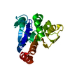
Mass: 18.015 Da / Num. of mol.: 41 / Source method: isolated from a natural source / Formula: H2O

-
Experimental details

-
Experiment

ExperimentMethod: X-RAY DIFFRACTION / Number of used crystals: 1

-
Sample preparation

CrystalDensity Matthews: 3.19 Å3/Da / Density % sol: 61.08 %
Crystal growTemperature: 295 K / Method: evaporation / pH: 6.5
Details: sodium acetate, EVAPORATION, temperature 295K, pH 4-9, pH 6.5

-
Data collection

DiffractionMean temperature: 100 K
Diffraction sourceSource: ROTATING ANODE / Type: RIGAKU / Wavelength: 1.5418 Å
DetectorType: RIGAKU RAXIS IV / Detector: IMAGE PLATE / Date: Oct 10, 2002
RadiationMonochromator: osmic mirror / Protocol: SINGLE WAVELENGTH / Monochromatic (M) / Laue (L): M / Scattering type: x-ray
Radiation wavelengthWavelength: 1.5418 Å / Relative weight: 1
ReflectionResolution: 2.5→20 Å / Num. all: 8696 / Num. obs: 8696 / % possible obs: 99.6 % / Observed criterion σ(F): 0 / Observed criterion σ(I): 0 / Biso Wilson estimate: 31.6 Å2
Reflection shellResolution: 2.5→2.59 Å / % possible all: 99.1

-
Processing

Software
NameVersionClassification
CNS1.1refinement
SCALEPACKdata scaling
CNSphasing
RefinementMethod to determine structure: MOLECULAR REPLACEMENT / Resolution: 2.5→19.84 Å / Rfactor Rfree error: 0.008 / Data cutoff high absF: 298114.11 / Data cutoff low absF: 0 / Isotropic thermal model: RESTRAINED / Cross valid method: THROUGHOUT / σ(F): 0
RfactorNum. reflection% reflectionSelection details
Rfree0.249 930 10.7 %RANDOM
Rwork0.228 ---
obs0.228 8683 99.5 %-
Solvent computationSolvent model: FLAT MODEL / Bsol: 39.4767 Å2 / ksol: 0.382065 e/Å3
Displacement parametersBiso mean: 27.9 Å2
Baniso -1Baniso -2Baniso -3
1-2.81 Å24.48 Å20 Å2
2--4.36 Å20 Å2
3----7.17 Å2
Refine analyze
FreeObs
Luzzati coordinate error0.35 Å0.31 Å
Luzzati d res low-5 Å
Luzzati sigma a0.29 Å0.3 Å
Refinement stepCycle: LAST / Resolution: 2.5→19.84 Å
ProteinNucleic acidLigandSolventTotal
Num. atoms1330 0 0 41 1371
Refine LS restraints
Refine-IDTypeDev idealDev ideal target
X-RAY DIFFRACTIONc_bond_d0.01
X-RAY DIFFRACTIONc_angle_deg1.4
X-RAY DIFFRACTIONc_dihedral_angle_d24.1
X-RAY DIFFRACTIONc_improper_angle_d1
X-RAY DIFFRACTIONc_mcbond_it01.5
X-RAY DIFFRACTIONc_mcangle_it02
X-RAY DIFFRACTIONc_scbond_it02
X-RAY DIFFRACTIONc_scangle_it02.5
LS refinement shellResolution: 2.5→2.66 Å / Rfactor Rfree error: 0.023 / Total num. of bins used: 6
RfactorNum. reflection% reflection
Rfree0.263 130 9.6 %
Rwork0.268 1231 -
obs--95.3 %
Xplor file
Refine-IDSerial noParam fileTopol file
X-RAY DIFFRACTION1PROTEIN_REP.PARAMPROTEIN.TOP
X-RAY DIFFRACTION2WATER_REP.PARAM

+
About Yorodumi

-
News

-
Feb 9, 2022. New format data for meta-information of EMDB entries

New format data for meta-information of EMDB entries

  • Version 3 of the EMDB header file is now the official format.
  • The previous official version 1.9 will be removed from the archive.

Related info.:EMDB header

External links:wwPDB to switch to version 3 of the EMDB data model

-
Aug 12, 2020. Covid-19 info

Covid-19 info

URL: https://pdbj.org/emnavi/covid19.php

New page: Covid-19 featured information page in EM Navigator.

Related info.:Covid-19 info / Mar 5, 2020. Novel coronavirus structure data

+
Mar 5, 2020. Novel coronavirus structure data

Novel coronavirus structure data

Related info.:Yorodumi Speices / Aug 12, 2020. Covid-19 info

External links:COVID-19 featured content - PDBj / Molecule of the Month (242):Coronavirus Proteases

+
Jan 31, 2019. EMDB accession codes are about to change! (news from PDBe EMDB page)

EMDB accession codes are about to change! (news from PDBe EMDB page)

  • The allocation of 4 digits for EMDB accession codes will soon come to an end. Whilst these codes will remain in use, new EMDB accession codes will include an additional digit and will expand incrementally as the available range of codes is exhausted. The current 4-digit format prefixed with “EMD-” (i.e. EMD-XXXX) will advance to a 5-digit format (i.e. EMD-XXXXX), and so on. It is currently estimated that the 4-digit codes will be depleted around Spring 2019, at which point the 5-digit format will come into force.
  • The EM Navigator/Yorodumi systems omit the EMD- prefix.

Related info.:Q: What is EMD? / ID/Accession-code notation in Yorodumi/EM Navigator

External links:EMDB Accession Codes are Changing Soon! / Contact to PDBj

+
Jul 12, 2017. Major update of PDB

Major update of PDB

  • wwPDB released updated PDB data conforming to the new PDBx/mmCIF dictionary.
  • This is a major update changing the version number from 4 to 5, and with Remediation, in which all the entries are updated.
  • In this update, many items about electron microscopy experimental information are reorganized (e.g. em_software).
  • Now, EM Navigator and Yorodumi are based on the updated data.

External links:wwPDB Remediation / Enriched Model Files Conforming to OneDep Data Standards Now Available in the PDB FTP Archive

-
Yorodumi

Thousand views of thousand structures

  • Yorodumi is a browser for structure data from EMDB, PDB, SASBDB, etc.
  • This page is also the successor to EM Navigator detail page, and also detail information page/front-end page for Omokage search.
  • The word "yorodu" (or yorozu) is an old Japanese word meaning "ten thousand". "mi" (miru) is to see.

Related info.:EMDB / PDB / SASBDB / Comparison of 3 databanks / Yorodumi Search / Aug 31, 2016. New EM Navigator & Yorodumi / Yorodumi Papers / Jmol/JSmol / Function and homology information / Changes in new EM Navigator and Yorodumi

Read more