 Movie
Movie Controller

[English] 日本語
 Yorodumi
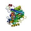
- PDB-1pbg: THE THREE-DIMENSIONAL STRUCTURE OF 6-PHOSPHO-BETA GALACTOSIDASE F... -
+ Open data

- Basic information
| Entry | Database: PDB / ID: 1pbg | ||||||
|---|---|---|---|---|---|---|---|
| Title | THE THREE-DIMENSIONAL STRUCTURE OF 6-PHOSPHO-BETA GALACTOSIDASE FROM LACTOCOCCUS LACTIS | ||||||
 Components Components | 6-PHOSPHO-BETA-D-GALACTOSIDASE | ||||||
 Keywords Keywords | HYDROLASE (GLYCOSYL HYDROLASE) | ||||||
| Function / homology |  Function and homology information Function and homology information6-phospho-beta-galactosidase / 6-phospho-beta-galactosidase activity / lactose catabolic process via tagatose-6-phosphate / beta-glucosidase activity / cytosol Similarity search - Function | ||||||
| Biological species |  Lactococcus lactis (lactic acid bacteria) Lactococcus lactis (lactic acid bacteria) | ||||||
| Method |  X-RAY DIFFRACTION / Resolution: 2.3 Å X-RAY DIFFRACTION / Resolution: 2.3 Å | ||||||
 Authors Authors | Wiesmann, C. / Schulz, G.E. | ||||||
 Citation Citation | Journal: Structure / Year: 1995 Title: The three-dimensional structure of 6-phospho-beta-galactosidase from Lactococcus lactis. Authors: Wiesmann, C. / Beste, G. / Hengstenberg, W. / Schulz, G.E. #1: Journal: Eur.J.Biochem. / Year: 1995Title: Identification of the Active-Site Nucleophile in 6-Phospho-Beta Galactosidase from Staphylococcus Aureus by Labelling with Synthetic Inhibitors Authors: Staedtler, P. / Hoenig, S. / Frank, R. / Withers, S.G. / Hengstenberg, W. #2: Journal: Protein Eng. / Year: 1993Title: 6-Phospho-Beta-Galactosidases of Gram-Positive and 6-Phospho-Beta-Glucosidase B of Gram-Negative Bacteria: Comparison of Structure and Function by Kinetic and Immunological Methods and ...Title: 6-Phospho-Beta-Galactosidases of Gram-Positive and 6-Phospho-Beta-Glucosidase B of Gram-Negative Bacteria: Comparison of Structure and Function by Kinetic and Immunological Methods and Mutagenesis of the Lacg Gene of Staphylococcus Aureus Authors: Witt, E. / Frank, R. / Hengstenberg, W. | ||||||
| History |
|
- Structure visualization
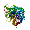
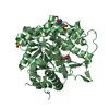
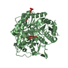
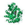
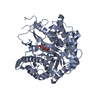
| Structure viewer | Molecule: MolmilJmol/JSmol |
|---|
- Downloads & links
-Download
| PDBx/mmCIF format | 1pbg.cif.gz | 200.1 KB | Display | PDBx/mmCIF format |
|---|---|---|---|---|
| PDB format | pdb1pbg.ent.gz | 161.3 KB | Display | PDB format |
| PDBx/mmJSON format | 1pbg.json.gz | Tree view | PDBx/mmJSON format | |
| Others |  Other downloads Other downloads |
-Validation report
| Arichive directory | https://data.pdbj.org/pub/pdb/validation_reports/pb/1pbgftp://data.pdbj.org/pub/pdb/validation_reports/pb/1pbg | HTTPS FTP |
|---|
-Related structure data
| Similar structure data |
|---|


-Links
 PDBj
PDBj- Assembly
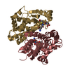
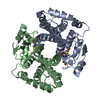
| Deposited unit | 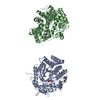
| ||||||||
|---|---|---|---|---|---|---|---|---|---|
| 1 | 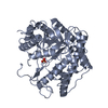
| ||||||||
| 2 | 
| ||||||||
| Unit cell |
| ||||||||
| Atom site foot note | 1: CIS PROLINE - PRO A 48 / 2: CIS PROLINE - PRO A 176 / 3: TRP A 421 - SER A 422 IS A CIS PEPTIDE. / 4: CIS PROLINE - PRO B 48 / 5: CIS PROLINE - PRO B 176 / 6: TRP B 421 - SER B 422 IS A CIS PEPTIDE. | ||||||||
| Noncrystallographic symmetry (NCS) | NCS oper: (Code: given Matrix: (0.999948, -0.00719, 0.00717), Vector: |
-Components
| #1: Protein | Mass: 54139.113 Da / Num. of mol.: 2 Source method: isolated from a genetically manipulated source Details: PRECIPITANT POLYETHYLENE GLYCOL Source: (gene. exp.) Lactococcus lactis (lactic acid bacteria)Strain: SUBSP. LACTIS 712 / Description: DELTA H DELTA 1TRP / Gene: LACG / Plasmid: PNZ316 / Gene (production host): LACG / Production host: #2: Chemical |   Mass: 96.063 Da / Num. of mol.: 2 / Source method: obtained synthetically / Formula: SO4 Mass: 96.063 Da / Num. of mol.: 2 / Source method: obtained synthetically / Formula: SO4#3: Water | ChemComp-HOH / |  Mass: 18.015 Da / Num. of mol.: 416 / Source method: isolated from a natural source / Formula: H2O Mass: 18.015 Da / Num. of mol.: 416 / Source method: isolated from a natural source / Formula: H2OCompound details | SECONDARY STRUCTURE ASSIGNMENT WAS DONE BY THE PROGRAM DSSP. FOR A MANUAL ASSIGNMENT CHECK THE MOST ...SECONDARY STRUCTURE ASSIGNMENT | |
|---|
-Experimental details
-Experiment
| Experiment | Method: X-RAY DIFFRACTION / Number of used crystals: 1 |
|---|
- Sample preparation
| Crystal | Density Matthews: 2.45 Å3/Da / Density % sol: 49.81 % | ||||||||||||||||||||||||||||||
|---|---|---|---|---|---|---|---|---|---|---|---|---|---|---|---|---|---|---|---|---|---|---|---|---|---|---|---|---|---|---|---|
| Crystal | *PLUS Density % sol: 50 % | ||||||||||||||||||||||||||||||
| Crystal grow | *PLUS Method: vapor diffusion, hanging drop / PH range low: 8 / PH range high: 6.7 | ||||||||||||||||||||||||||||||
| Components of the solutions | *PLUS
|
-Data collection
| Diffraction source | Wavelength: 1.5418 Å |
|---|---|
| Detector | Type: SIEMENS-NICOLET X100 / Detector: AREA DETECTOR / Date: 1994 |
| Radiation | Monochromatic (M) / Laue (L): M / Scattering type: x-ray |
| Radiation wavelength | Wavelength: 1.5418 Å / Relative weight: 1 |
| Reflection | Resolution: 2.3→100 Å / Num. obs: 36765 / % possible obs: 78.8 % / Redundancy: 3 % / Rmerge(I) obs: 0.069 |
| Reflection | *PLUS Observed criterion σ(I): 0 / Rmerge(I) obs: 0.069 |
- Processing
| Software |
| ||||||||||||||||||||||||||||||||||||||||||||||||||||||||||||||||||||||||||||||||
|---|---|---|---|---|---|---|---|---|---|---|---|---|---|---|---|---|---|---|---|---|---|---|---|---|---|---|---|---|---|---|---|---|---|---|---|---|---|---|---|---|---|---|---|---|---|---|---|---|---|---|---|---|---|---|---|---|---|---|---|---|---|---|---|---|---|---|---|---|---|---|---|---|---|---|---|---|---|---|---|---|---|
| Refinement | Resolution: 2.3→10 Å / σ(F): 0
| ||||||||||||||||||||||||||||||||||||||||||||||||||||||||||||||||||||||||||||||||
| Displacement parameters | Biso mean: 19.7 Å2 | ||||||||||||||||||||||||||||||||||||||||||||||||||||||||||||||||||||||||||||||||
| Refine analyze | Luzzati coordinate error obs: 0.23 Å | ||||||||||||||||||||||||||||||||||||||||||||||||||||||||||||||||||||||||||||||||
| Refinement step | Cycle: LAST / Resolution: 2.3→10 Å
| ||||||||||||||||||||||||||||||||||||||||||||||||||||||||||||||||||||||||||||||||
| Refine LS restraints |
| ||||||||||||||||||||||||||||||||||||||||||||||||||||||||||||||||||||||||||||||||
| Software | *PLUS Name: X-PLOR / Classification: refinement | ||||||||||||||||||||||||||||||||||||||||||||||||||||||||||||||||||||||||||||||||
| Refinement | *PLUS | ||||||||||||||||||||||||||||||||||||||||||||||||||||||||||||||||||||||||||||||||
| Solvent computation | *PLUS | ||||||||||||||||||||||||||||||||||||||||||||||||||||||||||||||||||||||||||||||||
| Displacement parameters | *PLUS | ||||||||||||||||||||||||||||||||||||||||||||||||||||||||||||||||||||||||||||||||
| Refine LS restraints | *PLUS
|
