 Movie
Movie Controller


+ Open data

- Basic information
| Entry | Database: PDB / ID: 1ooi | ||||||
|---|---|---|---|---|---|---|---|
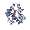
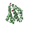
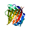
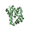
| Title | Crystal structure of LUSH from Drosophila melanogaster at pH 6.5 | ||||||
 Components Components | odorant binding protein LUSH | ||||||
 Keywords Keywords | TRANSPORT PROTEIN / LUSH / Alcohol / Odorant-Binding Protein | ||||||
| Function / homology |  Function and homology information Function and homology informationdiphenyl phthalate binding / dibutyl phthalate binding / response to pheromone / courtship behavior / pheromone binding / olfactory behavior / odorant binding / sensory perception of smell / response to ethanol / extracellular region Similarity search - Function | ||||||
| Biological species |  | ||||||
| Method |  X-RAY DIFFRACTION / MOLECULAR REPLACEMENT / Resolution: 2.04 Å X-RAY DIFFRACTION / MOLECULAR REPLACEMENT / Resolution: 2.04 Å | ||||||
 Authors Authors | Kruse, S.W. / Zhao, R. / Smith, D.P. / Jones, D.N.M. | ||||||
 Citation Citation | Journal: Nat.Struct.Biol. / Year: 2003 Title: Structure of a specific alcohol-binding site defined by the odorant binding protein LUSH from Drosophila melanogaster Authors: Kruse, S.W. / Zhao, R. / Smith, D.P. / Jones, D.N.M. | ||||||
| History |
|
- Structure visualization
| Structure viewer | Molecule: MolmilJmol/JSmol |
|---|
- Downloads & links
-Download
| PDBx/mmCIF format | 1ooi.cif.gz | 38.1 KB | Display | PDBx/mmCIF format |
|---|---|---|---|---|
| PDB format | pdb1ooi.ent.gz | 26.2 KB | Display | PDB format |
| PDBx/mmJSON format | 1ooi.json.gz | Tree view | PDBx/mmJSON format | |
| Others |  Other downloads Other downloads |
-Validation report
| Arichive directory | https://data.pdbj.org/pub/pdb/validation_reports/oo/1ooiftp://data.pdbj.org/pub/pdb/validation_reports/oo/1ooi | HTTPS FTP |
|---|
-Related structure data
| Related structure data |  1oofSC 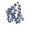 1oogC  1oohC S: Starting model for refinement C: citing same article ( |
|---|---|
| Similar structure data |


-Links
 PDBj
PDBj- Assembly
| Deposited unit | 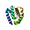
| ||||||||
|---|---|---|---|---|---|---|---|---|---|
| 1 |
| ||||||||
| Unit cell |
|
-Components
| #1: Protein | Mass: 14215.508 Da / Num. of mol.: 1 Source method: isolated from a genetically manipulated source Source: (gene. exp.)  |
|---|---|
| #2: Water | ChemComp-HOH /  Mass: 18.015 Da / Num. of mol.: 90 / Source method: isolated from a natural source / Formula: H2O Mass: 18.015 Da / Num. of mol.: 90 / Source method: isolated from a natural source / Formula: H2O |
| Has protein modification | Y |
-Experimental details
-Experiment
| Experiment | Method: X-RAY DIFFRACTION / Number of used crystals: 1 |
|---|
- Sample preparation
| Crystal | Density Matthews: 2.08 Å3/Da / Density % sol: 40.94 % | ||||||||||||||||||||||||||||||||||||
|---|---|---|---|---|---|---|---|---|---|---|---|---|---|---|---|---|---|---|---|---|---|---|---|---|---|---|---|---|---|---|---|---|---|---|---|---|---|
| Crystal grow | Temperature: 291 K / Method: vapor diffusion, hanging drop / pH: 6.5 Details: PEG 4000, MOPS, n-butanol, pH 6.5, VAPOR DIFFUSION, HANGING DROP, temperature 291K | ||||||||||||||||||||||||||||||||||||
| Crystal grow | *PLUS pH: 8 / Method: vapor diffusion | ||||||||||||||||||||||||||||||||||||
| Components of the solutions | *PLUS
|
-Data collection
| Diffraction | Mean temperature: 93 K |
|---|---|
| Diffraction source | Source: ROTATING ANODE / Type: RIGAKU RUH3R / Wavelength: 1.5418 Å |
| Detector | Type: RIGAKU RAXIS IV / Detector: IMAGE PLATE / Date: May 23, 2002 / Details: osmic blue optics |
| Radiation | Monochromator: NONE / Protocol: SINGLE WAVELENGTH / Monochromatic (M) / Laue (L): M / Scattering type: x-ray |
| Radiation wavelength | Wavelength: 1.5418 Å / Relative weight: 1 |
| Reflection | Resolution: 2.04→40 Å / Num. all: 7504 / Num. obs: 7504 / % possible obs: 100 % / Observed criterion σ(I): -3 / Redundancy: 14.9 % / Rmerge(I) obs: 0.054 / Net I/σ(I): 57.9 |
| Reflection shell | Resolution: 2.04→2.11 Å / Redundancy: 14.6 % / Rmerge(I) obs: 0.247 / Mean I/σ(I) obs: 10.7 / Num. unique all: 728 / % possible all: 100 |
| Reflection | *PLUS Num. measured all: 112114 |
| Reflection shell | *PLUS % possible obs: 100 % |
- Processing
| Software |
| |||||||||||||||||||||||||||||||||||||||||||||||||||||||||||||||||||||||||||
|---|---|---|---|---|---|---|---|---|---|---|---|---|---|---|---|---|---|---|---|---|---|---|---|---|---|---|---|---|---|---|---|---|---|---|---|---|---|---|---|---|---|---|---|---|---|---|---|---|---|---|---|---|---|---|---|---|---|---|---|---|---|---|---|---|---|---|---|---|---|---|---|---|---|---|---|---|
| Refinement | Method to determine structure: MOLECULAR REPLACEMENT Starting model: PDB ENTRY 1OOF Resolution: 2.04→40 Å / Cor.coef. Fo:Fc: 0.941 / Cor.coef. Fo:Fc free: 0.909 / Isotropic thermal model: isotropic temperature factors / Cross valid method: THROUGHOUT / σ(F): 0 / ESU R: 0.282 / ESU R Free: 0.211 / Stereochemistry target values: MAXIMUM LIKELIHOOD
| |||||||||||||||||||||||||||||||||||||||||||||||||||||||||||||||||||||||||||
| Solvent computation | Ion probe radii: 0.8 Å / Shrinkage radii: 0.8 Å / VDW probe radii: 1.4 Å / Solvent model: BABINET MODEL WITH MASK | |||||||||||||||||||||||||||||||||||||||||||||||||||||||||||||||||||||||||||
| Displacement parameters | Biso mean: 27.792 Å2
| |||||||||||||||||||||||||||||||||||||||||||||||||||||||||||||||||||||||||||
| Refinement step | Cycle: LAST / Resolution: 2.04→40 Å
| |||||||||||||||||||||||||||||||||||||||||||||||||||||||||||||||||||||||||||
| Refine LS restraints |
| |||||||||||||||||||||||||||||||||||||||||||||||||||||||||||||||||||||||||||
| LS refinement shell | Resolution: 2.04→2.15 Å / Total num. of bins used: 20
| |||||||||||||||||||||||||||||||||||||||||||||||||||||||||||||||||||||||||||
| Software | *PLUS Version: 5 / Classification: refinement | |||||||||||||||||||||||||||||||||||||||||||||||||||||||||||||||||||||||||||
| Refinement | *PLUS % reflection Rfree: 10 % / Rfactor Rfree: 0.247 / Rfactor Rwork: 0.195 | |||||||||||||||||||||||||||||||||||||||||||||||||||||||||||||||||||||||||||
| Solvent computation | *PLUS | |||||||||||||||||||||||||||||||||||||||||||||||||||||||||||||||||||||||||||
| Displacement parameters | *PLUS | |||||||||||||||||||||||||||||||||||||||||||||||||||||||||||||||||||||||||||
| Refine LS restraints | *PLUS
| |||||||||||||||||||||||||||||||||||||||||||||||||||||||||||||||||||||||||||
| LS refinement shell | *PLUS Rfactor Rwork: 0.209 |
