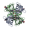
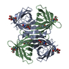
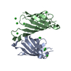
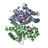
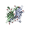
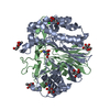
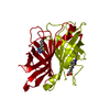
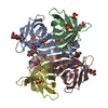
登録情報 データベース : PDB / ID : 1nqnタイトル Structure of Avm-W110K (W110K mutant of avidin) Avidin キーワード / / / / 機能・相同性 分子機能 ドメイン・相同性 構成要素
/ / / / / / / / / / / / / / / / / / / / / / 生物種 Gallus gallus (ニワトリ)手法 / / / 解像度 : 1.8 Å データ登録者 Pazy, Y. / Eisenberg-Domovich, Y. / Laitinen, O.H. / Kulomaa, M.S. / Bayer, E.A. / Wilchek, M. / Livnah, O. ジャーナル : J.Bacteriol. / 年 : 2003タイトル : Dimer-Tetramer Transition between Solution and Crystalline States of Streptavidin and Avidin Mutants.著者 : Pazy, Y. / Eisenberg-Domovich, Y. / Laitinen, O.H. / Kulomaa, M.S. / Bayer, E.A. / Wilchek, M. / Livnah, O. 履歴 登録 2003年1月22日 登録サイト / 処理サイト 改定 1.0 2003年7月15日 Provider / タイプ 改定 1.1 2008年4月29日 Group 改定 1.2 2011年7月13日 Group 改定 1.3 2018年1月31日 Group / カテゴリ / Item 改定 1.4 2021年10月27日 Group / Database referencesカテゴリ / pdbx_database_remark / struct_ref_seq_difItem _database_2.pdbx_DOI / _database_2.pdbx_database_accession ... _database_2.pdbx_DOI / _database_2.pdbx_database_accession / _pdbx_database_remark.text / _struct_ref_seq_dif.details 改定 1.5 2024年10月9日 Group / Structure summaryカテゴリ chem_comp_atom / chem_comp_bond ... chem_comp_atom / chem_comp_bond / pdbx_entry_details / pdbx_modification_feature
すべて表示 表示を減らす Remark 999 SEQUENCE According to Swiss-Prot entry P02701 there is a variant in residue 58 Ile -> Thr (IN APPR. ... SEQUENCE According to Swiss-Prot entry P02701 there is a variant in residue 58 Ile -> Thr (IN APPR. 50% OF THE CHAINS).
 ムービー
ムービー コントローラー


 データを開く
 基本情報
基本情報 要素
要素 キーワード
キーワード 機能・相同性情報
 X線回折 /
  データ登録者
 引用
引用 構造の表示
構造の表示 ダウンロードとリンク
 その他のダウンロード


 PDBj
PDBj 集合体
集合体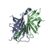


 分子量: 18.015 Da / 分子数: 94 / 由来タイプ: 天然 / 式: H2O
 試料調製
試料調製 / ビームライン: ID14-1 / 波長: 0.93 / 波長: 0.93 Å
 解析
解析