 Movie
Movie Controller

[English] 日本語
 Yorodumi
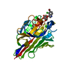
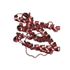
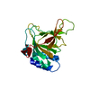
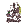
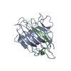
- PDB-1nln: CRYSTAL STRUCTURE OF HUMAN ADENOVIRUS 2 PROTEINASE WITH ITS 11 AM... -
+ Open data

- Basic information
| Entry | Database: PDB / ID: 1nln | ||||||
|---|---|---|---|---|---|---|---|
| Title | CRYSTAL STRUCTURE OF HUMAN ADENOVIRUS 2 PROTEINASE WITH ITS 11 AMINO ACID COFACTOR AT 1.6 ANGSTROM RESOLUTION | ||||||
 Components Components |
| ||||||
 Keywords Keywords | HYDROLASE / THIOL HYDROLASE / VIRAL PROTEINASE / PEPTIDE COFACTOR | ||||||
| Function / homology |  Function and homology information Function and homology informationnuclear capsid assembly / adenain / lysis of host organelle involved in viral entry into host cell / viral procapsid / microtubule-dependent intracellular transport of viral material towards nucleus / viral release from host cell / cysteine-type peptidase activity / virion component / viral capsid / host cell ...nuclear capsid assembly / adenain / lysis of host organelle involved in viral entry into host cell / viral procapsid / microtubule-dependent intracellular transport of viral material towards nucleus / viral release from host cell / cysteine-type peptidase activity / virion component / viral capsid / host cell / host cell cytoplasm / cysteine-type endopeptidase activity / host cell nucleus / proteolysis / DNA binding Similarity search - Function | ||||||
| Biological species |   Human adenovirus 2 Human adenovirus 2 | ||||||
| Method |  X-RAY DIFFRACTION / SYNCHROTRON / MOLECULAR REPLACEMENT / Resolution: 1.6 Å X-RAY DIFFRACTION / SYNCHROTRON / MOLECULAR REPLACEMENT / Resolution: 1.6 Å | ||||||
 Authors Authors | McGrath, W.J. / Ding, J. / Sweet, R.M. / Mangel, W.F. | ||||||
 Citation Citation | Journal: Biochim.Biophys.Acta / Year: 2003 Title: Crystallographic structure at 1.6-A resolution of the human adenovirus proteinase in a covalent complex with its 11-amino-acid peptide cofactor: insights on a new fold Authors: McGrath, W.J. / Ding, J. / Didwania, A. / Sweet, R.M. / Mangel, W.F. #1: Journal: Embo J. / Year: 1996Title: Crystal Structure of the Human Adenovirus Proteinase with its 11 Amino-acid Cofactor Authors: Ding, J. / McGrath, W.J. / Sweet, R.M. / Mangel, W.F. #2: Journal: J.Biol.Chem. / Year: 1996Title: Characterization of Three Components of Human Adenovirus Proteinase Activity in vitro #3: Journal: Nature / Year: 1993Title: Viral DNA and a Viral Peptide can act as Cofactors of Adenovirus Virion Proteinase Activity | ||||||
| History |
|
- Structure visualization
| Structure viewer | Molecule: MolmilJmol/JSmol |
|---|
- Downloads & links
-Download
| PDBx/mmCIF format | 1nln.cif.gz | 62.3 KB | Display | PDBx/mmCIF format |
|---|---|---|---|---|
| PDB format | pdb1nln.ent.gz | 44.7 KB | Display | PDB format |
| PDBx/mmJSON format | 1nln.json.gz | Tree view | PDBx/mmJSON format | |
| Others |  Other downloads Other downloads |
-Validation report
| Summary document | 1nln_validation.pdf.gz | 452.3 KB | Display | wwPDB validaton report |
|---|---|---|---|---|
| Full document | 1nln_full_validation.pdf.gz | 456.8 KB | Display | |
| Data in XML | 1nln_validation.xml.gz | 13.7 KB | Display | |
| Data in CIF | 1nln_validation.cif.gz | 19.7 KB | Display | |
| Arichive directory | https://data.pdbj.org/pub/pdb/validation_reports/nl/1nlnftp://data.pdbj.org/pub/pdb/validation_reports/nl/1nln | HTTPS FTP |
-Related structure data
| Related structure data |  1avpS S: Starting model for refinement |
|---|---|
| Similar structure data |


-Links
 PDBj
PDBj


- Assembly
| Deposited unit | 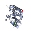
| ||||||||
|---|---|---|---|---|---|---|---|---|---|
| 1 |
| ||||||||
| Unit cell |
|
-Components
| #1: Protein | Mass: 23114.350 Da / Num. of mol.: 1 / Fragment: adenovirus proteinase Source method: isolated from a genetically manipulated source Source: (gene. exp.) Human adenovirus 2 / Genus: Mastadenovirus / Species: Human adenovirus C / Gene: L3-23k / Plasmid: pET13 / Species (production host): Escherichia coli / Production host:  |
|---|---|
| #2: Protein/peptide | Mass: 1353.640 Da / Num. of mol.: 1 / Fragment: pvic peptide / Source method: obtained synthetically Details: chemically synthesized C-terminal 11 amino acids from human adenovirus serotype 2 pVI molecule References: UniProt: P03274*PLUS |
| #3: Chemical | ChemComp-ACY /   Mass: 60.052 Da / Num. of mol.: 1 / Source method: obtained synthetically / Formula: C2H4O2 Mass: 60.052 Da / Num. of mol.: 1 / Source method: obtained synthetically / Formula: C2H4O2 |
| #4: Water | ChemComp-HOH /  Mass: 18.015 Da / Num. of mol.: 238 / Source method: isolated from a natural source / Formula: H2O Mass: 18.015 Da / Num. of mol.: 238 / Source method: isolated from a natural source / Formula: H2O |
-Experimental details
-Experiment
| Experiment | Method: X-RAY DIFFRACTION / Number of used crystals: 1 |
|---|
- Sample preparation
| Crystal | Density Matthews: 3.73 Å3/Da / Density % sol: 67.03 % |
|---|---|
| Crystal grow | Temperature: 298 K / Method: vapor diffusion, hanging drop / pH: 6.5 Details: cacodylate, sodium acetate, pH 6.5, VAPOR DIFFUSION, HANGING DROP, temperature 298K |
| Crystal grow | *PLUS Details: McGrath, W.J., (1996) J. Struct. Biol., 117, 77. |
-Data collection
| Diffraction | Mean temperature: 100 K |
|---|---|
| Diffraction source | Source: SYNCHROTRON / Site: NSLS  / Beamline: X12C / Wavelength: 1.15 Å / Beamline: X12C / Wavelength: 1.15 Å |
| Detector | Type: MARRESEARCH / Detector: AREA DETECTOR / Date: Aug 10, 1995 / Details: collimator |
| Radiation | Monochromator: Si crystal / Protocol: SINGLE WAVELENGTH / Monochromatic (M) / Laue (L): M / Scattering type: x-ray |
| Radiation wavelength | Wavelength: 1.15 Å / Relative weight: 1 |
| Reflection | Resolution: 1.6→30 Å / Num. obs: 44449 / % possible obs: 92.5 % / Observed criterion σ(F): 2 / Observed criterion σ(I): 2 / Redundancy: 11 % / Biso Wilson estimate: 16.3 Å2 / Rmerge(I) obs: 0.045 / Rsym value: 0.045 / Net I/σ(I): 30.6 |
| Reflection shell | Resolution: 1.6→50 Å / Redundancy: 5.4 % / Rmerge(I) obs: 0.166 / Mean I/σ(I) obs: 7.7 / Num. unique all: 3674 / Rsym value: 0.154 / % possible all: 77.1 |
| Reflection | *PLUS Num. obs: 44447 / Num. measured all: 166648 |
| Reflection shell | *PLUS Highest resolution: 1.6 Å / Lowest resolution: 1.66 Å / % possible obs: 77.1 % / Mean I/σ(I) obs: 7.6 |
- Processing
| Software |
| ||||||||||||||||||||
|---|---|---|---|---|---|---|---|---|---|---|---|---|---|---|---|---|---|---|---|---|---|
| Refinement | Method to determine structure: MOLECULAR REPLACEMENT Starting model: PDB Entry 1AVP Resolution: 1.6→30 Å / Isotropic thermal model: isotropic / Cross valid method: THROUGHOUT / σ(F): 2 / σ(I): 2 / Stereochemistry target values: Engh & Huber
| ||||||||||||||||||||
| Displacement parameters | Biso mean: 14.1 Å2 | ||||||||||||||||||||
| Refine analyze |
| ||||||||||||||||||||
| Refinement step | Cycle: LAST / Resolution: 1.6→30 Å
| ||||||||||||||||||||
| Refine LS restraints |
| ||||||||||||||||||||
| Software | *PLUS Name: X-PLOR / Classification: refinement | ||||||||||||||||||||
| Refine LS restraints | *PLUS
| ||||||||||||||||||||
| LS refinement shell | *PLUS Highest resolution: 1.6 Å / Lowest resolution: 1.66 Å |
