[English] 日本語
Yorodumi
- PDB-1nci: STRUCTURAL BASIS OF CELL-CELL ADHESION BY CADHERINS -

+
Open data


ID or keywords:

Loading...

-
Basic information

Entry
Database: PDB / ID: 1nci
TitleSTRUCTURAL BASIS OF CELL-CELL ADHESION BY CADHERINS
ComponentsN-CADHERINCadherin-2
KeywordsCELL ADHESION PROTEIN / CADHERIN
Function / homology
Function and homology information


mesenchymal cell migration / regulation of oligodendrocyte progenitor proliferation / regulation of postsynaptic density protein 95 clustering / radial glial cell differentiation / neuroligin clustering involved in postsynaptic membrane assembly / positive regulation of synaptic vesicle clustering / Regulation of Insulin-like Growth Factor (IGF) transport and uptake by Insulin-like Growth Factor Binding Proteins (IGFBPs) / Post-translational protein phosphorylation / Adherens junctions interactions / desmosome ...mesenchymal cell migration / regulation of oligodendrocyte progenitor proliferation / regulation of postsynaptic density protein 95 clustering / radial glial cell differentiation / neuroligin clustering involved in postsynaptic membrane assembly / positive regulation of synaptic vesicle clustering / Regulation of Insulin-like Growth Factor (IGF) transport and uptake by Insulin-like Growth Factor Binding Proteins (IGFBPs) / Post-translational protein phosphorylation / Adherens junctions interactions / desmosome / synaptic vesicle clustering / neural crest cell development / gamma-catenin binding / glial cell differentiation / cell-cell adhesion mediated by cadherin / telencephalon development / neuroepithelial cell differentiation / type B pancreatic cell development / neuronal stem cell population maintenance / alpha-catenin binding / fascia adherens / apicolateral plasma membrane / calcium-dependent cell-cell adhesion via plasma membrane cell adhesion molecules / Myogenesis / regulation of Rho protein signal transduction / postsynaptic specialization membrane / brain morphogenesis / catenin complex / cell-cell junction assembly / adherens junction organization / blood vessel morphogenesis / regulation of myelination / heterophilic cell-cell adhesion via plasma membrane cell adhesion molecules / regulation of axonogenesis / cortical actin cytoskeleton / nitric-oxide synthase binding / homophilic cell adhesion via plasma membrane adhesion molecules / plasma membrane raft / homeostasis of number of cells / intercalated disc / presynaptic active zone membrane / regulation of synaptic transmission, glutamatergic / synapse assembly / striated muscle cell differentiation / T-tubule / protein tyrosine kinase binding / protein localization to plasma membrane / adherens junction / brain development / negative regulation of canonical Wnt signaling pathway / sarcolemma / modulation of chemical synaptic transmission / cell morphogenesis / cell-cell adhesion / cerebral cortex development / beta-catenin binding / regulation of protein localization / cell-cell junction / cell migration / apical part of cell / lamellipodium / cell junction / basolateral plasma membrane / protein phosphatase binding / postsynaptic density / positive regulation of MAPK cascade / cell adhesion / neuron projection / cadherin binding / apical plasma membrane / glutamatergic synapse / synapse / calcium ion binding / protein-containing complex binding / protein kinase binding / enzyme binding / cell surface / protein-containing complex / RNA binding / membrane / identical protein binding / plasma membrane / cytoplasm
Similarity search - Function
Cadherin prodomain like / Cadherin prodomain / Cadherin prodomain like / Cadherin, Y-type LIR-motif / Cadherin, Y-type LIR-motif / Catenin binding domain superfamily / Cadherins / Cadherin / Cadherin conserved site / Cadherin domain signature. ...Cadherin prodomain like / Cadherin prodomain / Cadherin prodomain like / Cadherin, Y-type LIR-motif / Cadherin, Y-type LIR-motif / Catenin binding domain superfamily / Cadherins / Cadherin / Cadherin conserved site / Cadherin domain signature. / Cadherin repeats. / Cadherin domain / Cadherins domain profile. / Cadherin-like / Cadherin-like superfamily / Immunoglobulin-like / Sandwich / Mainly Beta
Similarity search - Domain/homology
URANYL (VI) ION / Cadherin-2
Similarity search - Component
Biological speciesMus musculus (house mouse)
MethodX-RAY DIFFRACTION / SYNCHROTRON / Resolution: 2.1 Å
AuthorsShapiro, L. / Fannon, A.M. / Kwong, P.D. / Thompson, A. / Lehmann, M.S. / Grubel, G. / Legrand, J.-F. / Als-Nielsen, J. / Colman, D.R. / Hendrickson, W.A.
CitationJournal: Nature / Year: 1995
Title: Structural basis of cell-cell adhesion by cadherins.
Authors: Shapiro, L. / Fannon, A.M. / Kwong, P.D. / Thompson, A. / Lehmann, M.S. / Grubel, G. / Legrand, J.F. / Als-Nielsen, J. / Colman, D.R. / Hendrickson, W.A.
History
DepositionMar 23, 1995Processing site: BNL
Revision 1.0Jul 10, 1995Provider: repository / Type: Initial release
Revision 1.1Mar 3, 2008Group: Version format compliance
Revision 1.2Jul 13, 2011Group: Version format compliance
Revision 1.3Nov 16, 2011Group: Atomic model
Revision 1.4Feb 14, 2024Group: Data collection / Database references ...Data collection / Database references / Derived calculations / Other
Category: chem_comp_atom / chem_comp_bond ...chem_comp_atom / chem_comp_bond / database_2 / pdbx_database_status / pdbx_struct_special_symmetry / struct_ref_seq_dif / struct_site
Item: _database_2.pdbx_DOI / _database_2.pdbx_database_accession ..._database_2.pdbx_DOI / _database_2.pdbx_database_accession / _pdbx_database_status.process_site / _struct_ref_seq_dif.details / _struct_site.pdbx_auth_asym_id / _struct_site.pdbx_auth_comp_id / _struct_site.pdbx_auth_seq_id

-
Structure visualization

Structure viewerMolecule:
MolmilJmol/JSmol

Downloads & links

-
Assembly

Deposited unit
A: N-CADHERIN
B: N-CADHERIN
hetero molecules


Theoretical massNumber of molelcules
Total (without water)24,7603
Polymers24,4902
Non-polymers2701
Water5,675315
1


  • Idetical with deposited unit
  • defined by author&software
TypeNameSymmetry operationNumber
identity operation1_555x,y,z1
Buried area2030 Å2
ΔGint-13 kcal/mol
Surface area10790 Å2
MethodPISA
Unit cell
Length a, b, c (Å)75.200, 57.500, 52.500
Angle α, β, γ (deg.)90.00, 90.00, 90.00
Int Tables number18
Space group name H-MP21212
Atom site foot note1: CIS PROLINE - PRO A 16 / 2: CIS PROLINE - PRO A 18 / 3: CIS PROLINE - PRO A 47 / 4: CIS PROLINE - PRO B 16 / 5: CIS PROLINE - PRO B 18 / 6: CIS PROLINE - PRO B 47
Components on special symmetry positions
IDModelComponents
11A-440-

HOH

21A-463-

HOH

31B-244-

HOH

41B-251-

HOH

Details
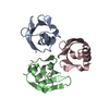
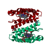
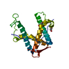
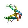
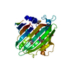
THE ASYMMETRIC UNIT (MOLECULES A AND B) COMPRISE A CADHERIN STRAND DIMER. AN ADHESION DIMER IS FORMED, FOR EXAMPLE, BETWEEN MOLECULE A AND THE (X + 1/2, 3/2 - Y, -1 -Z) SYMMETRY MATE OF MOLECULE B. SYMMETRY THE CRYSTALLOGRAPHIC SYMMETRY TRANSFORMATIONS PRESENTED BELOW GENERATE THE SUBUNITS OF THE POLYMERIC MOLECULE. APPLIED TO RESIDUES: B 1 .. 100 ADHESION DIMER MATE OF MOLECULE A IS A SYMMETRY-MATE OF MOLECULE B, AND CAN BE GENERATED BY THE FOLLOWING TRANSFORMATION: SYMMETRY1 1 1.000000 0.000000 0.000000 37.60000 SYMMETRY2 1 0.000000 1.500000 0.000000 86.25000 SYMMETRY3 1 0.000000 0.000000 1.000000 -52.50000

-
Components

#1: Protein N-CADHERIN / Cadherin-2


Mass: 12244.817 Da / Num. of mol.: 2
Source method: isolated from a genetically manipulated source
Source: (gene. exp.) Mus musculus (house mouse) / Plasmid: PGEX-2T / Production host: Escherichia coli (E. coli) / References: UniProt: P15116
#2: Chemical ChemComp-IUM / URANYL (VI) ION / Uranyl


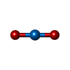
Mass: 270.028 Da / Num. of mol.: 1 / Source method: obtained synthetically / Formula: O2U
#3: Water ChemComp-HOH / water / Water


Mass: 18.015 Da / Num. of mol.: 315 / Source method: isolated from a natural source / Formula: H2O
Nonpolymer detailsTHE MODEL INCLUDES ONE URANYL ION (UO2 2+) BOUND AT THE PUTATIVE CALCIUM BINDING SITE.
Sequence detailsRESIDUE NUMBERING IS FOR THE MATURE PROTEIN.

-
Experimental details

-
Experiment

ExperimentMethod: X-RAY DIFFRACTION / Number of used crystals: 1

-
Sample preparation

CrystalDensity Matthews: 2.32 Å3/Da / Density % sol: 46.87 %
Description: 5 WAVELENGTH MAD EXPERIMENT; DATA COLLECTED NEAR URANIUM L(III) EDGE
Crystal grow
*PLUS
Temperature: 19.5 ℃ / pH: 4 / Method: vapor diffusion, hanging drop
Components of the solutions
*PLUS
IDConc.Common nameCrystal-IDSol-ID
135 mg/mlprotein1drop
20.5-0.6 Maqueous sodium sulfate1reservoir
30.1 MNa acetate1reservoir
45 mMUO2 acetate1reservoir

-
Data collection

Diffraction sourceSource: SYNCHROTRON / Site: NSLS / Beamline: X4A / Wavelength: 0.7217 - 0.7263
DetectorType: FUJI / Detector: IMAGE PLATE / Date: Aug 1, 1994
RadiationScattering type: x-ray
Radiation wavelength
IDWavelength (Å)Relative weight
10.72171
20.72631
ReflectionNum. obs: 33075 / % possible obs: 95.7 % / Observed criterion σ(I): 2 / Rmerge(I) obs: 0.038
Reflection
*PLUS
Highest resolution: 1.9 Å / Lowest resolution: 15 Å / Rmerge(I) obs: 0.038

-
Processing

Software
NameClassification
X-PLORmodel building
X-PLORrefinement
DENZOdata reduction
X-PLORphasing
RefinementResolution: 2.1→5 Å / σ(F): 2
RfactorNum. reflection% reflection
Rfree0.253 --
Rwork0.195 --
obs0.195 31010 96.2 %
Displacement parametersBiso mean: 17.1 Å2
Refinement stepCycle: LAST / Resolution: 2.1→5 Å
ProteinNucleic acidLigandSolventTotal
Num. atoms1557 0 3 315 1875
Refine LS restraints
Refine-IDTypeDev ideal
X-RAY DIFFRACTIONx_bond_d0.013
X-RAY DIFFRACTIONx_bond_d_na
X-RAY DIFFRACTIONx_bond_d_prot
X-RAY DIFFRACTIONx_angle_d
X-RAY DIFFRACTIONx_angle_d_na
X-RAY DIFFRACTIONx_angle_d_prot
X-RAY DIFFRACTIONx_angle_deg1.81
X-RAY DIFFRACTIONx_angle_deg_na
X-RAY DIFFRACTIONx_angle_deg_prot
X-RAY DIFFRACTIONx_dihedral_angle_d
X-RAY DIFFRACTIONx_dihedral_angle_d_na
X-RAY DIFFRACTIONx_dihedral_angle_d_prot
X-RAY DIFFRACTIONx_improper_angle_d
X-RAY DIFFRACTIONx_improper_angle_d_na
X-RAY DIFFRACTIONx_improper_angle_d_prot
X-RAY DIFFRACTIONx_mcbond_it
X-RAY DIFFRACTIONx_mcangle_it
X-RAY DIFFRACTIONx_scbond_it
X-RAY DIFFRACTIONx_scangle_it
Software
*PLUS
Name: X-PLOR / Classification: refinement
Refinement
*PLUS
Solvent computation
*PLUS
Displacement parameters
*PLUS

+
About Yorodumi

-
News

-
Feb 9, 2022. New format data for meta-information of EMDB entries

New format data for meta-information of EMDB entries

  • Version 3 of the EMDB header file is now the official format.
  • The previous official version 1.9 will be removed from the archive.

Related info.:EMDB header

External links:wwPDB to switch to version 3 of the EMDB data model

-
Aug 12, 2020. Covid-19 info

Covid-19 info

URL: https://pdbj.org/emnavi/covid19.php

New page: Covid-19 featured information page in EM Navigator.

Related info.:Covid-19 info / Mar 5, 2020. Novel coronavirus structure data

+
Mar 5, 2020. Novel coronavirus structure data

Novel coronavirus structure data

Related info.:Yorodumi Speices / Aug 12, 2020. Covid-19 info

External links:COVID-19 featured content - PDBj / Molecule of the Month (242):Coronavirus Proteases

+
Jan 31, 2019. EMDB accession codes are about to change! (news from PDBe EMDB page)

EMDB accession codes are about to change! (news from PDBe EMDB page)

  • The allocation of 4 digits for EMDB accession codes will soon come to an end. Whilst these codes will remain in use, new EMDB accession codes will include an additional digit and will expand incrementally as the available range of codes is exhausted. The current 4-digit format prefixed with “EMD-” (i.e. EMD-XXXX) will advance to a 5-digit format (i.e. EMD-XXXXX), and so on. It is currently estimated that the 4-digit codes will be depleted around Spring 2019, at which point the 5-digit format will come into force.
  • The EM Navigator/Yorodumi systems omit the EMD- prefix.

Related info.:Q: What is EMD? / ID/Accession-code notation in Yorodumi/EM Navigator

External links:EMDB Accession Codes are Changing Soon! / Contact to PDBj

+
Jul 12, 2017. Major update of PDB

Major update of PDB

  • wwPDB released updated PDB data conforming to the new PDBx/mmCIF dictionary.
  • This is a major update changing the version number from 4 to 5, and with Remediation, in which all the entries are updated.
  • In this update, many items about electron microscopy experimental information are reorganized (e.g. em_software).
  • Now, EM Navigator and Yorodumi are based on the updated data.

External links:wwPDB Remediation / Enriched Model Files Conforming to OneDep Data Standards Now Available in the PDB FTP Archive

-
Yorodumi

Thousand views of thousand structures

  • Yorodumi is a browser for structure data from EMDB, PDB, SASBDB, etc.
  • This page is also the successor to EM Navigator detail page, and also detail information page/front-end page for Omokage search.
  • The word "yorodu" (or yorozu) is an old Japanese word meaning "ten thousand". "mi" (miru) is to see.

Related info.:EMDB / PDB / SASBDB / Comparison of 3 databanks / Yorodumi Search / Aug 31, 2016. New EM Navigator & Yorodumi / Yorodumi Papers / Jmol/JSmol / Function and homology information / Changes in new EM Navigator and Yorodumi

Read more