 Movie
Movie Controller

[English] 日本語
 Yorodumi
- PDB-1mvp: STRUCTURAL STUDIES OF THE RETROVIRAL PROTEINASE FROM AVIAN MYELOB... -
+ Open data

- Basic information
| Entry | Database: PDB / ID: 1mvp | ||||||
|---|---|---|---|---|---|---|---|
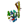
| Title | STRUCTURAL STUDIES OF THE RETROVIRAL PROTEINASE FROM AVIAN MYELOBLASTOSIS ASSOCIATED VIRUS | ||||||
 Components Components | MYELOBLASTOSIS ASSOCIATED VIRAL PROTEASE | ||||||
 Keywords Keywords | HYDROLASE(ASPARTIC PROTEINASE) | ||||||
| Function / homology |  Function and homology information Function and homology informationHydrolases; Acting on peptide bonds (peptidases); Aspartic endopeptidases / aspartic-type endopeptidase activity / proteolysis Similarity search - Function | ||||||
| Biological species |  Avian myeloblastosis-associated virus Avian myeloblastosis-associated virus | ||||||
| Method |  X-RAY DIFFRACTION / Resolution: 2.2 Å X-RAY DIFFRACTION / Resolution: 2.2 Å | ||||||
 Authors Authors | Ohlendorf, D.H. / Foundling, S.I. / Salemme, F.R. | ||||||
 Citation Citation | Journal: Proteins / Year: 1992 Title: Structural studies of the retroviral proteinase from avian myeloblastosis associated virus. Authors: Ohlendorf, D.H. / Foundling, S.I. / Wendoloski, J.J. / Sedlacek, J. / Strop, P. / Salemme, F.R. | ||||||
| History |
|
- Structure visualization
| Structure viewer | Molecule: MolmilJmol/JSmol |
|---|
- Downloads & links
-Download
| PDBx/mmCIF format | 1mvp.cif.gz | 56.5 KB | Display | PDBx/mmCIF format |
|---|---|---|---|---|
| PDB format | pdb1mvp.ent.gz | 41.9 KB | Display | PDB format |
| PDBx/mmJSON format | 1mvp.json.gz | Tree view | PDBx/mmJSON format | |
| Others |  Other downloads Other downloads |
-Validation report
| Arichive directory | https://data.pdbj.org/pub/pdb/validation_reports/mv/1mvpftp://data.pdbj.org/pub/pdb/validation_reports/mv/1mvp | HTTPS FTP |
|---|
-Related structure data
| Similar structure data |
|---|


-Links
 PDBj
PDBj- Assembly
| Deposited unit | 
| ||||||||
|---|---|---|---|---|---|---|---|---|---|
| 1 |
| ||||||||
| Unit cell |
| ||||||||
| Components on special symmetry positions |
| ||||||||
| Noncrystallographic symmetry (NCS) | NCS oper: (Code: given Matrix: (-0.06347, 0.57288, -0.81718), Vector: Details | THE TRANSFORMATION PRESENTED IN THE *MTRIX1* RECORDS BELOW WILL YIELD THE COORDINATES OF CHAIN *B* WHEN APPLIED TO THE COORDINATES OF CHAIN *A*. | |
-Components
| #1: Protein | Mass: 13611.836 Da / Num. of mol.: 2 Source method: isolated from a genetically manipulated source Source: (gene. exp.) Avian myeloblastosis-associated virus / Genus: AlpharetrovirusReferences: UniProt: P26315, Hydrolases; Acting on peptide bonds (peptidases); Aspartic endopeptidases #2: Water | ChemComp-HOH / |  Mass: 18.015 Da / Num. of mol.: 133 / Source method: isolated from a natural source / Formula: H2O Mass: 18.015 Da / Num. of mol.: 133 / Source method: isolated from a natural source / Formula: H2O |
|---|
-Experimental details
-Experiment
| Experiment | Method: X-RAY DIFFRACTION |
|---|
- Sample preparation
| Crystal | Density Matthews: 3.3 Å3/Da / Density % sol: 62.74 % | ||||||||||||||||||||||||||||||||||||||||||||||||||||||||||||
|---|---|---|---|---|---|---|---|---|---|---|---|---|---|---|---|---|---|---|---|---|---|---|---|---|---|---|---|---|---|---|---|---|---|---|---|---|---|---|---|---|---|---|---|---|---|---|---|---|---|---|---|---|---|---|---|---|---|---|---|---|---|
| Crystal grow | *PLUS pH: 5.6 / Method: vapor diffusion, hanging drop | ||||||||||||||||||||||||||||||||||||||||||||||||||||||||||||
| Components of the solutions | *PLUS
|
-Data collection
| Radiation | Scattering type: x-ray |
|---|---|
| Radiation wavelength | Relative weight: 1 |
| Reflection | *PLUS Highest resolution: 2.2 Å / Num. obs: 16968 / Num. measured all: 196542 |
- Processing
| Software | Name: PROLSQ / Classification: refinement | ||||||||||||||||||||||||||||||||||||||||||||||||||||||||||||||||||||||||||||||||||||
|---|---|---|---|---|---|---|---|---|---|---|---|---|---|---|---|---|---|---|---|---|---|---|---|---|---|---|---|---|---|---|---|---|---|---|---|---|---|---|---|---|---|---|---|---|---|---|---|---|---|---|---|---|---|---|---|---|---|---|---|---|---|---|---|---|---|---|---|---|---|---|---|---|---|---|---|---|---|---|---|---|---|---|---|---|---|
| Refinement | Resolution: 2.2→5 Å Details: THE SG ATOMS OF CYS A 113 AND CYS B 113 HAVE BEEN REFINED IN TWO ALTERNATE POSITIONS WITH AN OCCUPANCY OF 0.5 FOR EACH POSITION.
| ||||||||||||||||||||||||||||||||||||||||||||||||||||||||||||||||||||||||||||||||||||
| Refinement step | Cycle: LAST / Resolution: 2.2→5 Å
| ||||||||||||||||||||||||||||||||||||||||||||||||||||||||||||||||||||||||||||||||||||
| Refine LS restraints |
| ||||||||||||||||||||||||||||||||||||||||||||||||||||||||||||||||||||||||||||||||||||
| Software | *PLUS Name: PROLSQ / Classification: refinement | ||||||||||||||||||||||||||||||||||||||||||||||||||||||||||||||||||||||||||||||||||||
| Refinement | *PLUS Rfactor obs: 0.172 | ||||||||||||||||||||||||||||||||||||||||||||||||||||||||||||||||||||||||||||||||||||
| Solvent computation | *PLUS | ||||||||||||||||||||||||||||||||||||||||||||||||||||||||||||||||||||||||||||||||||||
| Displacement parameters | *PLUS |
