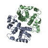
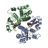
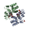
 Movie
Movie Controller


+ Open data

- Basic information
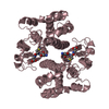
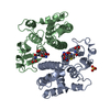
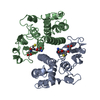
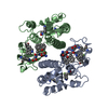
| Entry | Database: PDB / ID: 1mtc | ||||||
|---|---|---|---|---|---|---|---|
| Title | GLUTATHIONE TRANSFERASE MUTANT Y115F | ||||||
 Components Components | Glutathione S-transferase YB1 | ||||||
 Keywords Keywords | TRANSFERASE / GLUTATHIONE TRANSFERASE / PROTEIN DYNAMICS / PROTEIN CATALYSIS | ||||||
| Function / homology |  Function and homology information Function and homology informationGlutathione conjugation / nitrobenzene metabolic process / cellular detoxification of nitrogen compound / hepoxilin biosynthetic process / glutathione derivative biosynthetic process / glutathione binding / response to metal ion / prostaglandin metabolic process / nickel cation binding / glutathione transferase ...Glutathione conjugation / nitrobenzene metabolic process / cellular detoxification of nitrogen compound / hepoxilin biosynthetic process / glutathione derivative biosynthetic process / glutathione binding / response to metal ion / prostaglandin metabolic process / nickel cation binding / glutathione transferase / glutathione transferase activity / response to amino acid / xenobiotic catabolic process / response to axon injury / steroid binding / glutathione metabolic process / response to lead ion / cellular response to xenobiotic stimulus / sensory perception of smell / response to ethanol / protein kinase binding / enzyme binding / protein homodimerization activity / protein-containing complex / mitochondrion / extracellular region / identical protein binding / cytoplasm / cytosol Similarity search - Function | ||||||
| Biological species |  | ||||||
| Method |  X-RAY DIFFRACTION / MOLECULAR REPLACEMENT / Resolution: 2.2 Å X-RAY DIFFRACTION / MOLECULAR REPLACEMENT / Resolution: 2.2 Å | ||||||
 Authors Authors | Ladner, J.E. / Xiao, G. / Armstrong, R.N. / Gilliland, G.L. | ||||||
 Citation Citation | Journal: Biochemistry / Year: 2002 Title: Local protein dynamics and catalysis: detection of segmental motion associated with rate-limiting product release by a glutathione transferase Authors: Codreanu, S.G. / Ladner, J.E. / Xiao, G. / Stourman, N.V. / Hachey, D.L. / Gilliland, G.L. / Armstrong, R.N. | ||||||
| History |
|
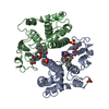
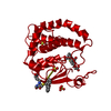
- Structure visualization
| Structure viewer | Molecule: MolmilJmol/JSmol |
|---|
- Downloads & links
-Download
| PDBx/mmCIF format | 1mtc.cif.gz | 106.8 KB | Display | PDBx/mmCIF format |
|---|---|---|---|---|
| PDB format | pdb1mtc.ent.gz | 82.1 KB | Display | PDB format |
| PDBx/mmJSON format | 1mtc.json.gz | Tree view | PDBx/mmJSON format | |
| Others |  Other downloads Other downloads |
-Validation report
| Arichive directory | https://data.pdbj.org/pub/pdb/validation_reports/mt/1mtcftp://data.pdbj.org/pub/pdb/validation_reports/mt/1mtc | HTTPS FTP |
|---|
-Related structure data
| Related structure data |  3gstS S: Starting model for refinement |
|---|---|
| Similar structure data |


-Links
 PDBj
PDBj

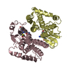
- Assembly
| Deposited unit | 
| ||||||||||
|---|---|---|---|---|---|---|---|---|---|---|---|
| 1 |
| ||||||||||
| Unit cell |
|
-Components
| #1: Protein | Mass: 25802.791 Da / Num. of mol.: 2 / Mutation: Y115F Source method: isolated from a genetically manipulated source Details: COMPLEX WITH (9R,10R)-9-(S-GLUTATHIONYL)-10-HYDROXY-9,10-DIHROPHENANTHRENE Source: (gene. exp.)  #2: Chemical |   Mass: 501.552 Da / Num. of mol.: 2 / Source method: obtained synthetically / Formula: C24H27N3O7S Mass: 501.552 Da / Num. of mol.: 2 / Source method: obtained synthetically / Formula: C24H27N3O7S#3: Water | ChemComp-HOH / |  Mass: 18.015 Da / Num. of mol.: 185 / Source method: isolated from a natural source / Formula: H2O Mass: 18.015 Da / Num. of mol.: 185 / Source method: isolated from a natural source / Formula: H2O |
|---|
-Experimental details
-Experiment
| Experiment | Method: X-RAY DIFFRACTION / Number of used crystals: 1 |
|---|
- Sample preparation
| Crystal | Density Matthews: 1.88 Å3/Da / Density % sol: 45.03 % | ||||||||||||||||||||||||||||||||||||||||||||||||||||||
|---|---|---|---|---|---|---|---|---|---|---|---|---|---|---|---|---|---|---|---|---|---|---|---|---|---|---|---|---|---|---|---|---|---|---|---|---|---|---|---|---|---|---|---|---|---|---|---|---|---|---|---|---|---|---|---|
| Crystal grow | Method: vapor diffusion, hanging drop / pH: 8 Details: ammonoium sulfate at pH 8.0, VAPOR DIFFUSION, HANGING DROP | ||||||||||||||||||||||||||||||||||||||||||||||||||||||
| Crystal grow | *PLUS Method: vapor diffusion, sitting drop / Details: Ji, X., (1992) Biochemistry, 31, 10169. | ||||||||||||||||||||||||||||||||||||||||||||||||||||||
| Components of the solutions | *PLUS
|
-Data collection
| Diffraction | Mean temperature: 143 K |
|---|---|
| Diffraction source | Source: ROTATING ANODE / Type: SIEMENS / Wavelength: 1.5418 Å |
| Detector | Type: BRUKER / Detector: AREA DETECTOR / Date: Mar 25, 1995 |
| Radiation | Protocol: SINGLE WAVELENGTH / Monochromatic (M) / Laue (L): M / Scattering type: x-ray |
| Radiation wavelength | Wavelength: 1.5418 Å / Relative weight: 1 |
| Reflection | Resolution: 2.2→10 Å / Num. obs: 21775 / % possible obs: 94 % / Redundancy: 3 % / Rmerge(I) obs: 0.11 / Net I/σ(I): 33 |
| Reflection shell | Resolution: 2.2→2.26 Å / Redundancy: 3 % / Rmerge(I) obs: 0.23 / Mean I/σ(I) obs: 8 / % possible all: 91 |
| Reflection shell | *PLUS % possible obs: 91 % |
- Processing
| Software |
| |||||||||||||||||||||||||||||||||
|---|---|---|---|---|---|---|---|---|---|---|---|---|---|---|---|---|---|---|---|---|---|---|---|---|---|---|---|---|---|---|---|---|---|---|
| Refinement | Method to determine structure: MOLECULAR REPLACEMENT Starting model: 3GST Resolution: 2.2→10 Å / Num. parameters: 15571 / Num. restraintsaints: 15303 / Cross valid method: FREE R / σ(F): 0 / Stereochemistry target values: ENGH & HUBER Details: ANISOTROPIC SCALING APPLIED BY THE METHOD OF PARKIN, MOEZZI & HOPE, J.APPL.CRYST.28(1995)53-56
| |||||||||||||||||||||||||||||||||
| Refine analyze | Num. disordered residues: 0 / Occupancy sum hydrogen: 0 / Occupancy sum non hydrogen: 3889 | |||||||||||||||||||||||||||||||||
| Refinement step | Cycle: LAST / Resolution: 2.2→10 Å
| |||||||||||||||||||||||||||||||||
| Refine LS restraints |
| |||||||||||||||||||||||||||||||||
| Software | *PLUS Name: SHELXL / Version: 97 / Classification: refinement | |||||||||||||||||||||||||||||||||
| Refinement | *PLUS Highest resolution: 2.2 Å / Lowest resolution: 10 Å / Rfactor Rwork: 0.203 | |||||||||||||||||||||||||||||||||
| Solvent computation | *PLUS | |||||||||||||||||||||||||||||||||
| Displacement parameters | *PLUS |
