 Movie
Movie Controller

[English] 日本語
 Yorodumi
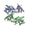
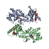
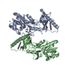
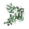
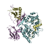
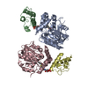
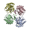
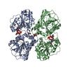
- PDB-1mrz: Crystal structure of a flavin binding protein from Thermotoga Mar... -
+ Open data

- Basic information
| Entry | Database: PDB / ID: 1mrz | ||||||
|---|---|---|---|---|---|---|---|
| Title | Crystal structure of a flavin binding protein from Thermotoga Maritima, TM379 | ||||||
 Components Components | Riboflavin kinase/FMN adenylyltransferase | ||||||
 Keywords Keywords | TRANSFERASE / Rossmann fold / flavin binding domain / 6-stranded beta barrel / nucleotide binding domain / Structural Genomics / BSGC structure funded by NIH / Protein Structure Initiative / PSI / Berkeley Structural Genomics Center | ||||||
| Function / homology |  Function and homology information Function and homology informationriboflavin metabolic process / riboflavin kinase / FAD synthase / FMN adenylyltransferase activity / FAD biosynthetic process / riboflavin kinase activity / FMN biosynthetic process / riboflavin biosynthetic process / ATP binding / metal ion binding Similarity search - Function | ||||||
| Biological species |   Thermotoga maritima (bacteria) Thermotoga maritima (bacteria) | ||||||
| Method |  X-RAY DIFFRACTION / SYNCHROTRON / MOLECULAR REPLACEMENT / Resolution: 1.9 Å X-RAY DIFFRACTION / SYNCHROTRON / MOLECULAR REPLACEMENT / Resolution: 1.9 Å | ||||||
 Authors Authors | Wang, W. / Kim, R. / Jancarik, J. / Yokota, H. / Kim, S.-H. / Berkeley Structural Genomics Center (BSGC) | ||||||
 Citation Citation | Journal: Proteins / Year: 2003 Title: Crystal structure of a flavin-binding protein from Thermotoga Maritima Authors: Wang, W. / Kim, R. / Jancarik, J. / Yokota, H. / Kim, S.-H. | ||||||
| History |
|
- Structure visualization
| Structure viewer | Molecule: MolmilJmol/JSmol |
|---|
- Downloads & links
-Download
| PDBx/mmCIF format | 1mrz.cif.gz | 128.5 KB | Display | PDBx/mmCIF format |
|---|---|---|---|---|
| PDB format | pdb1mrz.ent.gz | 99.7 KB | Display | PDB format |
| PDBx/mmJSON format | 1mrz.json.gz | Tree view | PDBx/mmJSON format | |
| Others |  Other downloads Other downloads |
-Validation report
| Arichive directory | https://data.pdbj.org/pub/pdb/validation_reports/mr/1mrzftp://data.pdbj.org/pub/pdb/validation_reports/mr/1mrz | HTTPS FTP |
|---|
-Related structure data
| Similar structure data | |
|---|---|
| Other databases |


-Links
 PDBj
PDBj- Assembly
| Deposited unit | 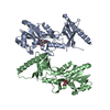
| ||||||||
|---|---|---|---|---|---|---|---|---|---|
| 1 |
| ||||||||
| Unit cell |
|
-Components
| #1: Protein | Mass: 33665.820 Da / Num. of mol.: 2 Source method: isolated from a genetically manipulated source Source: (gene. exp.) Thermotoga maritima (bacteria) / Gene: TM379 / Plasmid: pSJS1244 / Species (production host): Escherichia coli / Production host: #2: Chemical |   Mass: 192.124 Da / Num. of mol.: 2 / Source method: obtained synthetically / Formula: C6H8O7 Mass: 192.124 Da / Num. of mol.: 2 / Source method: obtained synthetically / Formula: C6H8O7#3: Water | ChemComp-HOH / |  Mass: 18.015 Da / Num. of mol.: 442 / Source method: isolated from a natural source / Formula: H2O Mass: 18.015 Da / Num. of mol.: 442 / Source method: isolated from a natural source / Formula: H2O |
|---|
-Experimental details
-Experiment
| Experiment | Method: X-RAY DIFFRACTION / Number of used crystals: 1 |
|---|
- Sample preparation
| Crystal | Density Matthews: 2.51 Å3/Da / Density % sol: 50.54 % | ||||||||||||||||||||||||||||||||||||||||||||||||||||||
|---|---|---|---|---|---|---|---|---|---|---|---|---|---|---|---|---|---|---|---|---|---|---|---|---|---|---|---|---|---|---|---|---|---|---|---|---|---|---|---|---|---|---|---|---|---|---|---|---|---|---|---|---|---|---|---|
| Crystal grow | Temperature: 293 K / Method: evaporation / pH: 7.5 Details: 0.2M Na Citrate, 23% PEG 3350, 10% Glycerol, pH 7.5, EVAPORATION, temperature 293K | ||||||||||||||||||||||||||||||||||||||||||||||||||||||
| Crystal grow | *PLUS Method: vapor diffusion, hanging drop | ||||||||||||||||||||||||||||||||||||||||||||||||||||||
| Components of the solutions | *PLUS
|
-Data collection
| Diffraction | Mean temperature: 100 K |
|---|---|
| Diffraction source | Source: SYNCHROTRON / Site: ALS  / Beamline: 5.0.2 / Wavelength: 0.97911 Å / Beamline: 5.0.2 / Wavelength: 0.97911 Å |
| Detector | Type: ADSC QUANTUM 4 / Detector: CCD / Date: May 2, 2001 |
| Radiation | Protocol: SINGLE WAVELENGTH / Monochromatic (M) / Laue (L): M / Scattering type: x-ray |
| Radiation wavelength | Wavelength: 0.97911 Å / Relative weight: 1 |
| Reflection | Resolution: 1.9→25 Å / Num. obs: 52473 / % possible obs: 96.9 % / Observed criterion σ(F): 1 / Observed criterion σ(I): 1 / Redundancy: 3.9 % / Rsym value: 0.038 / Net I/σ(I): 27 |
| Reflection shell | Resolution: 1.9→1.93 Å / Mean I/σ(I) obs: 4.1 / Num. unique all: 2670 / Rsym value: 0.299 / % possible all: 82.2 |
| Reflection | *PLUS Highest resolution: 1.9 Å / Lowest resolution: 30 Å / Num. obs: 51627 / Redundancy: 3.8 % / Rmerge(I) obs: 0.038 |
| Reflection shell | *PLUS Highest resolution: 1.9 Å / Lowest resolution: 1.93 Å / % possible obs: 82.2 % / Redundancy: 3.5 % / Num. unique obs: 2190 / Rmerge(I) obs: 0.299 / Mean I/σ(I) obs: 3 |
- Processing
| Software |
| ||||||||||||||||||||||||||||||||||||||||||||||||||||||||||||||||||||||||||||||||||||||||||||||||||||||||||||||||||||||||||||||||||
|---|---|---|---|---|---|---|---|---|---|---|---|---|---|---|---|---|---|---|---|---|---|---|---|---|---|---|---|---|---|---|---|---|---|---|---|---|---|---|---|---|---|---|---|---|---|---|---|---|---|---|---|---|---|---|---|---|---|---|---|---|---|---|---|---|---|---|---|---|---|---|---|---|---|---|---|---|---|---|---|---|---|---|---|---|---|---|---|---|---|---|---|---|---|---|---|---|---|---|---|---|---|---|---|---|---|---|---|---|---|---|---|---|---|---|---|---|---|---|---|---|---|---|---|---|---|---|---|---|---|---|---|
| Refinement | Method to determine structure: MOLECULAR REPLACEMENT Starting model: MAD Resolution: 1.9→20 Å / Cor.coef. Fo:Fc: 0.942 / Cor.coef. Fo:Fc free: 0.933 / SU B: 3.446 / SU ML: 0.104 / TLS residual ADP flag: LIKELY RESIDUAL / Cross valid method: THROUGHOUT / σ(F): 0 / ESU R: 0.182 / ESU R Free: 0.15 / Stereochemistry target values: MAXIMUM LIKELIHOOD
| ||||||||||||||||||||||||||||||||||||||||||||||||||||||||||||||||||||||||||||||||||||||||||||||||||||||||||||||||||||||||||||||||||
| Solvent computation | Ion probe radii: 0.8 Å / Shrinkage radii: 0.8 Å / VDW probe radii: 1.4 Å / Solvent model: BABINET MODEL WITH MASK | ||||||||||||||||||||||||||||||||||||||||||||||||||||||||||||||||||||||||||||||||||||||||||||||||||||||||||||||||||||||||||||||||||
| Displacement parameters | Biso mean: 21.308 Å2
| ||||||||||||||||||||||||||||||||||||||||||||||||||||||||||||||||||||||||||||||||||||||||||||||||||||||||||||||||||||||||||||||||||
| Refinement step | Cycle: LAST / Resolution: 1.9→20 Å
| ||||||||||||||||||||||||||||||||||||||||||||||||||||||||||||||||||||||||||||||||||||||||||||||||||||||||||||||||||||||||||||||||||
| Refine LS restraints |
| ||||||||||||||||||||||||||||||||||||||||||||||||||||||||||||||||||||||||||||||||||||||||||||||||||||||||||||||||||||||||||||||||||
| LS refinement shell | Resolution: 1.9→1.949 Å / Total num. of bins used: 20 /
| ||||||||||||||||||||||||||||||||||||||||||||||||||||||||||||||||||||||||||||||||||||||||||||||||||||||||||||||||||||||||||||||||||
| Refinement TLS params. | Method: refined / Refine-ID: X-RAY DIFFRACTION
| ||||||||||||||||||||||||||||||||||||||||||||||||||||||||||||||||||||||||||||||||||||||||||||||||||||||||||||||||||||||||||||||||||
| Refinement TLS group |
| ||||||||||||||||||||||||||||||||||||||||||||||||||||||||||||||||||||||||||||||||||||||||||||||||||||||||||||||||||||||||||||||||||
| Software | *PLUS Version: 5 / Classification: refinement | ||||||||||||||||||||||||||||||||||||||||||||||||||||||||||||||||||||||||||||||||||||||||||||||||||||||||||||||||||||||||||||||||||
| Refinement | *PLUS Highest resolution: 1.9 Å / Lowest resolution: 30 Å / Rfactor Rfree: 0.233 / Rfactor Rwork: 0.216 | ||||||||||||||||||||||||||||||||||||||||||||||||||||||||||||||||||||||||||||||||||||||||||||||||||||||||||||||||||||||||||||||||||
| Solvent computation | *PLUS | ||||||||||||||||||||||||||||||||||||||||||||||||||||||||||||||||||||||||||||||||||||||||||||||||||||||||||||||||||||||||||||||||||
| Displacement parameters | *PLUS | ||||||||||||||||||||||||||||||||||||||||||||||||||||||||||||||||||||||||||||||||||||||||||||||||||||||||||||||||||||||||||||||||||
| Refine LS restraints | *PLUS
| ||||||||||||||||||||||||||||||||||||||||||||||||||||||||||||||||||||||||||||||||||||||||||||||||||||||||||||||||||||||||||||||||||
| LS refinement shell | *PLUS Highest resolution: 1.9 Å / Lowest resolution: 1.93 Å |
