 Movie
Movie Controller

[English] 日本語
 Yorodumi
- PDB-1m9a: Crystal structure of the 26 kDa glutathione S-transferase from Sc... -
+ Open data

- Basic information
| Entry | Database: PDB / ID: 1m9a | ||||||
|---|---|---|---|---|---|---|---|
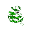
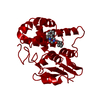
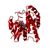
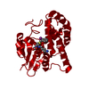
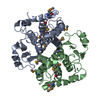
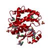
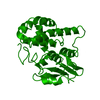
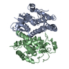
| Title | Crystal structure of the 26 kDa glutathione S-transferase from Schistosoma japonicum complexed with S-hexylglutathione | ||||||
 Components Components | Glutathione S-Transferase 26 kDa | ||||||
 Keywords Keywords | TRANSFERASE / Glutathione transferase / antigen / multigene family | ||||||
| Function / homology |  Function and homology information Function and homology informationglutathione transferase / glutathione transferase activity / glutathione metabolic process Similarity search - Function | ||||||
| Biological species |  | ||||||
| Method |  X-RAY DIFFRACTION / SYNCHROTRON / MOLECULAR REPLACEMENT / Resolution: 2.1 Å X-RAY DIFFRACTION / SYNCHROTRON / MOLECULAR REPLACEMENT / Resolution: 2.1 Å | ||||||
 Authors Authors | Cardoso, R.M.F. / Daniels, D.S. / Bruns, C.M. / Tainer, J.A. | ||||||
 Citation Citation | Journal: PROTEINS: STRUCT.,FUNCT.,GENET. / Year: 2003 Title: Characterization of the electrophile binding site and substrate binding mode of the 26-kDa glutathione S-transferase from Schistosoma japonicum Authors: Cardoso, R.M.F. / Daniels, D.S. / Bruns, C.M. / Tainer, J.A. #1: Journal: J.Mol.Biol. / Year: 1995Title: Crystal structures of a schistosomal drug and vaccine target: glutathione S-transferase from Schistosoma japonica and its complex with the leading antischistosomal drug praziquantel Authors: McTigue, M.A. / Williams, D.R. / Tainer, J.A. #2: Journal: Protein Sci. / Year: 1994Title: Three-dimensional structure of Schistosoma japonicum glutathione S-transferase fused with a six-amino acid conserved neutralizing epitope of gp41 from HIV Authors: Lim, K. / Ho, J.X. / Keeling, K. / Gilliland, G.L. / Ji, X. / Ruker, F. / Carter, D.C. | ||||||
| History |
|
- Structure visualization
| Structure viewer | Molecule: MolmilJmol/JSmol |
|---|
- Downloads & links
-Download
| PDBx/mmCIF format | 1m9a.cif.gz | 61.8 KB | Display | PDBx/mmCIF format |
|---|---|---|---|---|
| PDB format | pdb1m9a.ent.gz | 45.6 KB | Display | PDB format |
| PDBx/mmJSON format | 1m9a.json.gz | Tree view | PDBx/mmJSON format | |
| Others |  Other downloads Other downloads |
-Validation report
| Arichive directory | https://data.pdbj.org/pub/pdb/validation_reports/m9/1m9aftp://data.pdbj.org/pub/pdb/validation_reports/m9/1m9a | HTTPS FTP |
|---|
-Related structure data
| Related structure data |  1m99C  1m9bC  1gtaS S: Starting model for refinement C: citing same article ( |
|---|---|
| Similar structure data |

-Links
 PDBj
PDBj

- Assembly
| Deposited unit | 
| ||||||||||||
|---|---|---|---|---|---|---|---|---|---|---|---|---|---|
| 1 | 
| ||||||||||||
| Unit cell |
| ||||||||||||
| Components on special symmetry positions |
| ||||||||||||
| Details | There is one GST subunit per asymmetric unit and the second subunit of the GST dimer is generated by the symmetry operation: -x, -x+y, -z |
-Components
| #1: Protein | Mass: 25534.723 Da / Num. of mol.: 1 Source method: isolated from a genetically manipulated source Source: (gene. exp.)  |
|---|---|
| #2: Chemical | ChemComp-GTX /   Mass: 392.491 Da / Num. of mol.: 1 / Source method: obtained synthetically / Formula: C16H30N3O6S Mass: 392.491 Da / Num. of mol.: 1 / Source method: obtained synthetically / Formula: C16H30N3O6S |
| #3: Water | ChemComp-HOH /  Mass: 18.015 Da / Num. of mol.: 174 / Source method: isolated from a natural source / Formula: H2O Mass: 18.015 Da / Num. of mol.: 174 / Source method: isolated from a natural source / Formula: H2O |
-Experimental details
-Experiment
| Experiment | Method: X-RAY DIFFRACTION / Number of used crystals: 1 |
|---|
- Sample preparation
| Crystal | Density Matthews: 2.87 Å3/Da / Density % sol: 56.85 % | ||||||||||||||||||||||||||||||||||||
|---|---|---|---|---|---|---|---|---|---|---|---|---|---|---|---|---|---|---|---|---|---|---|---|---|---|---|---|---|---|---|---|---|---|---|---|---|---|
| Crystal grow | Temperature: 298 K / Method: vapor diffusion, hanging drop / pH: 5.6 Details: ammonium sulfate,ethanol, dithiothrietol, threalose, sodium acetate, pH 5.6, VAPOR DIFFUSION, HANGING DROP, temperature 298K | ||||||||||||||||||||||||||||||||||||
| Crystal grow | *PLUS | ||||||||||||||||||||||||||||||||||||
| Components of the solutions | *PLUS
|
-Data collection
| Diffraction | Mean temperature: 90 K |
|---|---|
| Diffraction source | Source: SYNCHROTRON / Site: SSRL  / Beamline: BL7-1 / Wavelength: 1.08 Å / Beamline: BL7-1 / Wavelength: 1.08 Å |
| Detector | Type: MARRESEARCH / Detector: IMAGE PLATE / Date: Dec 17, 1996 |
| Radiation | Monochromator: Si(111) / Protocol: SINGLE WAVELENGTH / Monochromatic (M) / Laue (L): M / Scattering type: x-ray |
| Radiation wavelength | Wavelength: 1.08 Å / Relative weight: 1 |
| Reflection | Resolution: 2.1→50 Å / Num. all: 17330 / Num. obs: 17330 / % possible obs: 94.4 % / Observed criterion σ(I): -3 / Redundancy: 4.6 % / Biso Wilson estimate: 19.1 Å2 / Rsym value: 0.062 / Net I/σ(I): 24 |
| Reflection shell | Resolution: 2.1→2.18 Å / Redundancy: 3.6 % / Mean I/σ(I) obs: 3.5 / Num. unique all: 1137 / Rsym value: 0.296 / % possible all: 63.4 |
| Reflection | *PLUS Lowest resolution: 27.9 Å / Num. measured all: 80211 / Rmerge(I) obs: 0.062 |
| Reflection shell | *PLUS % possible obs: 63.4 % / Rmerge(I) obs: 0.296 |
- Processing
| Software |
| ||||||||||||||||||||||||||||||||||||||||||||||||||||||||||||||||||||||||||||||||
|---|---|---|---|---|---|---|---|---|---|---|---|---|---|---|---|---|---|---|---|---|---|---|---|---|---|---|---|---|---|---|---|---|---|---|---|---|---|---|---|---|---|---|---|---|---|---|---|---|---|---|---|---|---|---|---|---|---|---|---|---|---|---|---|---|---|---|---|---|---|---|---|---|---|---|---|---|---|---|---|---|---|
| Refinement | Method to determine structure: MOLECULAR REPLACEMENT Starting model: PDB entry 1GTA Resolution: 2.1→33.9 Å / Rfactor Rfree error: 0.008 / Data cutoff high rms absF: 1957943.39 / Isotropic thermal model: RESTRAINED / Cross valid method: THROUGHOUT / σ(F): 0 / Stereochemistry target values: Engh & Huber
| ||||||||||||||||||||||||||||||||||||||||||||||||||||||||||||||||||||||||||||||||
| Solvent computation | Solvent model: FLAT MODEL / Bsol: 58.88 Å2 / ksol: 0.396569 e/Å3 | ||||||||||||||||||||||||||||||||||||||||||||||||||||||||||||||||||||||||||||||||
| Displacement parameters | Biso mean: 30.8 Å2
| ||||||||||||||||||||||||||||||||||||||||||||||||||||||||||||||||||||||||||||||||
| Refine analyze |
| ||||||||||||||||||||||||||||||||||||||||||||||||||||||||||||||||||||||||||||||||
| Refinement step | Cycle: LAST / Resolution: 2.1→33.9 Å
| ||||||||||||||||||||||||||||||||||||||||||||||||||||||||||||||||||||||||||||||||
| Refine LS restraints |
| ||||||||||||||||||||||||||||||||||||||||||||||||||||||||||||||||||||||||||||||||
| LS refinement shell | Resolution: 2.1→2.23 Å / Rfactor Rfree error: 0.028 / Total num. of bins used: 6
| ||||||||||||||||||||||||||||||||||||||||||||||||||||||||||||||||||||||||||||||||
| Xplor file |
| ||||||||||||||||||||||||||||||||||||||||||||||||||||||||||||||||||||||||||||||||
| Refinement | *PLUS Highest resolution: 2.1 Å / Lowest resolution: 27.9 Å / % reflection Rfree: 5 % / Rfactor Rwork: 0.2 | ||||||||||||||||||||||||||||||||||||||||||||||||||||||||||||||||||||||||||||||||
| Solvent computation | *PLUS | ||||||||||||||||||||||||||||||||||||||||||||||||||||||||||||||||||||||||||||||||
| Displacement parameters | *PLUS | ||||||||||||||||||||||||||||||||||||||||||||||||||||||||||||||||||||||||||||||||
| Refine LS restraints | *PLUS
|
