 Movie
Movie Controller


+ Open data

- Basic information
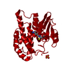
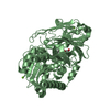
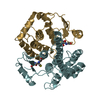
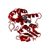
| Entry | Database: PDB / ID: 3cru | ||||||
|---|---|---|---|---|---|---|---|
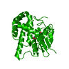
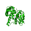
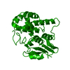
| Title | Structural characterization of an engineered allosteric protein | ||||||
 Components Components | Glutathione S-transferase class-mu 26 kDa isozyme | ||||||
 Keywords Keywords | TRANSFERASE / protein design / allosteric switch / pH-response | ||||||
| Function / homology |  Function and homology information Function and homology informationglutathione transferase / glutathione transferase activity / glutathione metabolic process Similarity search - Function | ||||||
| Biological species |  | ||||||
| Method |  X-RAY DIFFRACTION / MOLECULAR REPLACEMENT / Resolution: 2.3 Å X-RAY DIFFRACTION / MOLECULAR REPLACEMENT / Resolution: 2.3 Å | ||||||
 Authors Authors | Sagermann, M. / Chapleau, R. / DeLorimier, E. / Lei, M. | ||||||
 Citation Citation | Journal: Protein Sci. / Year: 2009 Title: Using affinity chromatography to engineer and characterize pH-dependent protein switches. Authors: Sagermann, M. / Chapleau, R.R. / DeLorimier, E. / Lei, M. | ||||||
| History |
|
- Structure visualization
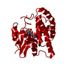
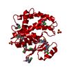
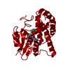
| Structure viewer | Molecule: MolmilJmol/JSmol |
|---|
- Downloads & links
-Download
| PDBx/mmCIF format | 3cru.cif.gz | 58.4 KB | Display | PDBx/mmCIF format |
|---|---|---|---|---|
| PDB format | pdb3cru.ent.gz | 42.4 KB | Display | PDB format |
| PDBx/mmJSON format | 3cru.json.gz | Tree view | PDBx/mmJSON format | |
| Others |  Other downloads Other downloads |
-Validation report
| Arichive directory | https://data.pdbj.org/pub/pdb/validation_reports/cr/3cruftp://data.pdbj.org/pub/pdb/validation_reports/cr/3cru | HTTPS FTP |
|---|
-Related structure data
| Related structure data |  3crtC 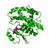 3d0zC 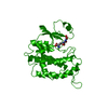 1gneS  3crs S: Starting model for refinement C: citing same article ( |
|---|---|
| Similar structure data |
-Links
 PDBj
PDBj

- Assembly
| Deposited unit | 
| ||||||||
|---|---|---|---|---|---|---|---|---|---|
| 1 | 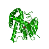
| ||||||||
| Unit cell |
|
-Components
| #1: Protein | Mass: 25063.148 Da / Num. of mol.: 1 / Mutation: L50C Source method: isolated from a genetically manipulated source Source: (gene. exp.)  |
|---|---|
| #2: Chemical | ChemComp-GSH /   Mass: 307.323 Da / Num. of mol.: 1 / Source method: obtained synthetically / Formula: C10H17N3O6S Mass: 307.323 Da / Num. of mol.: 1 / Source method: obtained synthetically / Formula: C10H17N3O6S |
| #3: Water | ChemComp-HOH /  Mass: 18.015 Da / Num. of mol.: 90 / Source method: isolated from a natural source / Formula: H2O Mass: 18.015 Da / Num. of mol.: 90 / Source method: isolated from a natural source / Formula: H2O |
-Experimental details
-Experiment
| Experiment | Method: X-RAY DIFFRACTION / Number of used crystals: 1 |
|---|
- Sample preparation
| Crystal | Density Matthews: 2.46 Å3/Da / Density % sol: 49.97 % |
|---|---|
| Crystal grow | Temperature: 298 K / Method: vapor diffusion, hanging drop / pH: 9.5 Details: 20% PEG8000, 50 mM Tris-HCL, 3 mM B-ME., pH 9.5, VAPOR DIFFUSION, HANGING DROP, temperature 298K |
-Data collection
| Diffraction | Mean temperature: 150 K |
|---|---|
| Diffraction source | Source: ROTATING ANODE / Type: RIGAKU FR-E+ SUPERBRIGHT / Wavelength: 1.5418 Å |
| Detector | Type: RIGAKU RAXIS IV++ / Detector: IMAGE PLATE / Date: Oct 4, 2007 / Details: Rigaku Varimax HR |
| Radiation | Monochromator: Rigaku VariMax HR optics / Protocol: SINGLE WAVELENGTH / Monochromatic (M) / Laue (L): M / Scattering type: x-ray |
| Radiation wavelength | Wavelength: 1.5418 Å / Relative weight: 1 |
| Reflection | Resolution: 2.3→19.507 Å / Num. all: 21386 / Num. obs: 17967 / % possible obs: 84 % / Observed criterion σ(F): 0 / Observed criterion σ(I): 0 / Redundancy: 1.99 % / Biso Wilson estimate: 29.96 Å2 / Rmerge(I) obs: 0.61 / Net I/σ(I): 14.87 |
| Reflection shell | Resolution: 2.3→3 Å / Redundancy: 1.91 % / Rmerge(I) obs: 0.158 / Mean I/σ(I) obs: 6.2 / Num. unique all: 8979 / % possible all: 76.5 |
- Processing
| Software |
| |||||||||||||||||||||||||
|---|---|---|---|---|---|---|---|---|---|---|---|---|---|---|---|---|---|---|---|---|---|---|---|---|---|---|
| Refinement | Method to determine structure: MOLECULAR REPLACEMENT Starting model: PDBid: 1GNE with the engineered peptide deleted. Resolution: 2.3→19.507 Å / Isotropic thermal model: Anisotropic / Cross valid method: THROUGHOUT / σ(F): 0 / σ(I): 0 / Stereochemistry target values: Engh & Huber Details: last four residues (HPPK) could not be modeled reliably and were omitted from the model.
| |||||||||||||||||||||||||
| Displacement parameters |
| |||||||||||||||||||||||||
| Refinement step | Cycle: LAST / Resolution: 2.3→19.507 Å
| |||||||||||||||||||||||||
| Refine LS restraints |
|
