 Movie
Movie Controller

[English] 日本語
 Yorodumi
- PDB-1lq2: Crystal structure of barley beta-D-glucan glucohydrolase isoenzym... -
+ Open data

- Basic information
| Entry | Database: PDB / ID: 1lq2 | |||||||||
|---|---|---|---|---|---|---|---|---|---|---|
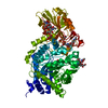
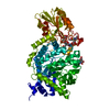
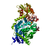
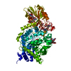
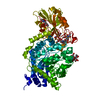
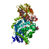
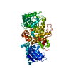
| Title | Crystal structure of barley beta-D-glucan glucohydrolase isoenzyme Exo1 in complex with gluco-phenylimidazole | |||||||||
 Components Components | Beta-D-glucan glucohydrolase isoenzyme Exo1 | |||||||||
 Keywords Keywords | HYDROLASE / 2-domain fold / ligand-protein complex | |||||||||
| Function / homology |  Function and homology information Function and homology informationbeta-glucosidase / hydrolase activity, hydrolyzing O-glycosyl compounds / carbohydrate metabolic process / extracellular region Similarity search - Function | |||||||||
| Biological species |  | |||||||||
| Method |  X-RAY DIFFRACTION / MOLECULAR REPLACEMENT / Resolution: 2.7 Å X-RAY DIFFRACTION / MOLECULAR REPLACEMENT / Resolution: 2.7 Å | |||||||||
 Authors Authors | Hrmova, M. / De Gori, R. / Smith, B.J. / Vasella, A. / Varghese, J.N. / Fincher, G.B. | |||||||||
 Citation Citation | Journal: J.Biol.Chem. / Year: 2004 Title: Three-dimensional Structure of the Barley {beta}-D-Glucan Glucohydrolase in Complex with a Transition State Mimic. Authors: Hrmova, M. / De Gori, R. / Smith, B.J. / Vasella, A. / Varghese, J.N. / Fincher, G.B. | |||||||||
| History |
| |||||||||
| Remark 999 | SEQUENCE The authors state there is an error in the cDNA sequencing of AF102868 (GenBank accession ...SEQUENCE The authors state there is an error in the cDNA sequencing of AF102868 (GenBank accession number). Residue 320 (sequence database residue 345) is a LYS and is not ASN. |
- Structure visualization
| Structure viewer | Molecule: MolmilJmol/JSmol |
|---|
- Downloads & links
-Download
| PDBx/mmCIF format | 1lq2.cif.gz | 136.8 KB | Display | PDBx/mmCIF format |
|---|---|---|---|---|
| PDB format | pdb1lq2.ent.gz | 104.2 KB | Display | PDB format |
| PDBx/mmJSON format | 1lq2.json.gz | Tree view | PDBx/mmJSON format | |
| Others |  Other downloads Other downloads |
-Validation report
| Summary document | 1lq2_validation.pdf.gz | 618 KB | Display | wwPDB validaton report |
|---|---|---|---|---|
| Full document | 1lq2_full_validation.pdf.gz | 634 KB | Display | |
| Data in XML | 1lq2_validation.xml.gz | 15.9 KB | Display | |
| Data in CIF | 1lq2_validation.cif.gz | 24.6 KB | Display | |
| Arichive directory | https://data.pdbj.org/pub/pdb/validation_reports/lq/1lq2ftp://data.pdbj.org/pub/pdb/validation_reports/lq/1lq2 | HTTPS FTP |
-Related structure data
| Related structure data |  1ieqS S: Starting model for refinement |
|---|---|
| Similar structure data |


-Links
 PDBj
PDBj- Assembly
| Deposited unit | 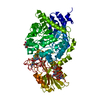
| ||||||||
|---|---|---|---|---|---|---|---|---|---|
| 1 |
| ||||||||
| Unit cell |
| ||||||||
| Details | The biological assembly is a monomer constructed from an (alpha/beta)8 barrel and an (alpha/beta)6 sandwich |
-Components
-Protein , 1 types, 1 molecules A
| #1: Protein | Mass: 65054.082 Da / Num. of mol.: 1 / Source method: isolated from a natural source / Source: (natural) References: GenBank: 4566505, UniProt: Q9XEI3*PLUS, glucan 1,3-beta-glucosidase |
|---|
-Sugars , 3 types, 3 molecules 
| #2: Polysaccharide | 2-acetamido-2-deoxy-beta-D-glucopyranose-(1-2)-alpha-D-mannopyranose-(1-6)-alpha-D-mannopyranose-(1- ...2-acetamido-2-deoxy-beta-D-glucopyranose-(1-2)-alpha-D-mannopyranose-(1-6)-alpha-D-mannopyranose-(1-4)-2-acetamido-2-deoxy-beta-D-glucopyranose-(1-4)-[alpha-L-fucopyranose-(1-3)]2-acetamido-2-deoxy-beta-D-glucopyranose Source method: isolated from a genetically manipulated source |
|---|---|
| #3: Polysaccharide | beta-D-mannopyranose-(1-4)-2-acetamido-2-deoxy-beta-D-glucopyranose-(1-4)-[alpha-L-fucopyranose-(1- ...beta-D-mannopyranose-(1-4)-2-acetamido-2-deoxy-beta-D-glucopyranose-(1-4)-[alpha-L-fucopyranose-(1-3)]2-acetamido-2-deoxy-beta-D-glucopyranose Source method: isolated from a genetically manipulated source |
| #4: Sugar | ChemComp-NAG /  Type: D-saccharide, beta linking / Mass: 221.208 Da / Num. of mol.: 1 Type: D-saccharide, beta linking / Mass: 221.208 Da / Num. of mol.: 1Source method: isolated from a genetically manipulated source Formula: C8H15NO6 |
-Non-polymers , 3 types, 243 molecules 


| #5: Chemical | ChemComp-IDD / ( Mass: 276.288 Da / Num. of mol.: 1 / Source method: obtained synthetically / Formula: C14H16N2O4 Mass: 276.288 Da / Num. of mol.: 1 / Source method: obtained synthetically / Formula: C14H16N2O4 |
|---|---|
| #6: Chemical | ChemComp-GOL /  Mass: 92.094 Da / Num. of mol.: 1 / Source method: obtained synthetically / Formula: C3H8O3 Mass: 92.094 Da / Num. of mol.: 1 / Source method: obtained synthetically / Formula: C3H8O3 |
| #7: Water | ChemComp-HOH / Mass: 18.015 Da / Num. of mol.: 241 / Source method: isolated from a natural source / Formula: H2O |
-Experimental details
-Experiment
| Experiment | Method: X-RAY DIFFRACTION / Number of used crystals: 1 |
|---|
- Sample preparation
| Crystal | Density Matthews: 3.56 Å3/Da / Density % sol: 65.42 % | ||||||||||||||||||||||||||||||||||||||||
|---|---|---|---|---|---|---|---|---|---|---|---|---|---|---|---|---|---|---|---|---|---|---|---|---|---|---|---|---|---|---|---|---|---|---|---|---|---|---|---|---|---|
| Crystal grow | Temperature: 277 K / Method: vapor diffusion, hanging drop / pH: 7 Details: Ammonium sulphate, PEG 400, Sodium acetate, Hepes-NaOH, pH 7.0, VAPOR DIFFUSION, HANGING DROP, temperature 277K | ||||||||||||||||||||||||||||||||||||||||
| Crystal grow | *PLUS Temperature: 277-279 K / Method: vapor diffusion, hanging drop / Details: Hrmova, M., (1998) Acta Cryst., D54, 687. | ||||||||||||||||||||||||||||||||||||||||
| Components of the solutions | *PLUS
|
-Data collection
| Diffraction | Mean temperature: 100 K |
|---|---|
| Diffraction source | Source: ROTATING ANODE / Type: MACSCIENCE / Wavelength: 1.5418 Å |
| Detector | Type: RIGAKU RAXIS IV / Detector: IMAGE PLATE / Date: Aug 15, 2001 / Details: Mono-capillary optics |
| Radiation | Protocol: SINGLE WAVELENGTH / Monochromatic (M) / Laue (L): M / Scattering type: x-ray |
| Radiation wavelength | Wavelength: 1.5418 Å / Relative weight: 1 |
| Reflection | Resolution: 2.62→50 Å / Num. obs: 24521 / % possible obs: 45.3 % / Observed criterion σ(F): 0 / Observed criterion σ(I): 0 / Redundancy: 1.81 % / Rmerge(I) obs: 0.13 / Net I/σ(I): 5.9 |
| Reflection shell | Resolution: 2.62→2.69 Å / Rmerge(I) obs: 0.582 / % possible all: 61.1 |
| Reflection | *PLUS Num. obs: 44625 / % possible obs: 73.6 % / Num. measured all: 70377 / Rmerge(I) obs: 0.13 |
| Reflection shell | *PLUS % possible obs: 45.4 % / Rmerge(I) obs: 0.583 |
- Processing
| Software |
| ||||||||||||||||||||
|---|---|---|---|---|---|---|---|---|---|---|---|---|---|---|---|---|---|---|---|---|---|
| Refinement | Method to determine structure: MOLECULAR REPLACEMENT Starting model: PDB Entry 1IEQ Resolution: 2.7→25 Å / Isotropic thermal model: Isotropic / Cross valid method: THROUGHOUT / σ(F): 0 / σ(I): 0 / Stereochemistry target values: Engh & Huber
| ||||||||||||||||||||
| Refine analyze | Luzzati coordinate error obs: 0.3 Å | ||||||||||||||||||||
| Refinement step | Cycle: LAST / Resolution: 2.7→25 Å
| ||||||||||||||||||||
| Refine LS restraints |
| ||||||||||||||||||||
| Refinement | *PLUS Highest resolution: 2.62 Å / % reflection Rfree: 5 % / Rfactor Rfree: 0.2729 / Rfactor Rwork: 0.2096 | ||||||||||||||||||||
| Solvent computation | *PLUS | ||||||||||||||||||||
| Displacement parameters | *PLUS |
