 Movie
Movie Controller

[English] 日本語
 Yorodumi
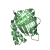
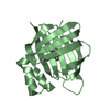
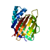
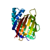
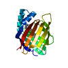
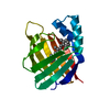
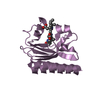
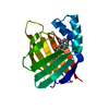
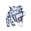
- PDB-1lba: THE STRUCTURE OF BACTERIOPHAGE T7 LYSOZYME, A ZINC AMIDASE AND AN... -
+ Open data

- Basic information
| Entry | Database: PDB / ID: 1lba | ||||||
|---|---|---|---|---|---|---|---|
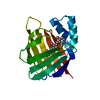
| Title | THE STRUCTURE OF BACTERIOPHAGE T7 LYSOZYME, A ZINC AMIDASE AND AN INHIBITOR OF T7 RNA POLYMERASE | ||||||
 Components Components | T7 LYSOZYME | ||||||
 Keywords Keywords | HYDROLASE(ACTING ON LINEAR AMIDES) | ||||||
| Function / homology |  Function and homology information Function and homology information N-acetylmuramoyl-L-alanine amidase / N-acetylmuramoyl-L-alanine amidase activity / negative regulation of viral transcription / viral release from host cell by cytolysis / peptidoglycan catabolic process / host cell cytoplasm / defense response to bacterium / zinc ion binding N-acetylmuramoyl-L-alanine amidase / N-acetylmuramoyl-L-alanine amidase activity / negative regulation of viral transcription / viral release from host cell by cytolysis / peptidoglycan catabolic process / host cell cytoplasm / defense response to bacterium / zinc ion bindingSimilarity search - Function | ||||||
| Biological species |   Enterobacteria phage T7 (virus) Enterobacteria phage T7 (virus) | ||||||
| Method | X-RAY DIFFRACTION / Resolution: 2.2 Å | ||||||
 Authors Authors | Cheng, X. | ||||||
 Citation Citation | Journal: Proc.Natl.Acad.Sci.USA / Year: 1994 Title: The structure of bacteriophage T7 lysozyme, a zinc amidase and an inhibitor of T7 RNA polymerase. Authors: Cheng, X. / Zhang, X. / Pflugrath, J.W. / Studier, F.W. | ||||||
| History |
| ||||||
| Remark 700 | SHEET BESIDES THE STRANDS REPRESENTED ON *S1* IN SHEET RECORDS BELOW, THERE ARE OTHER STRANDS IN ...SHEET BESIDES THE STRANDS REPRESENTED ON *S1* IN SHEET RECORDS BELOW, THERE ARE OTHER STRANDS IN THIS STRUCTURE WHICH DO NOT FORM ANY SHEETS. |
- Structure visualization
| Structure viewer | Molecule: MolmilJmol/JSmol |
|---|
- Downloads & links
-Download
| PDBx/mmCIF format | 1lba.cif.gz | 40.8 KB | Display | PDBx/mmCIF format |
|---|---|---|---|---|
| PDB format | pdb1lba.ent.gz | 28.1 KB | Display | PDB format |
| PDBx/mmJSON format | 1lba.json.gz | Tree view | PDBx/mmJSON format | |
| Others |  Other downloads Other downloads |
-Validation report
| Arichive directory | https://data.pdbj.org/pub/pdb/validation_reports/lb/1lbaftp://data.pdbj.org/pub/pdb/validation_reports/lb/1lba | HTTPS FTP |
|---|
-Related structure data
| Similar structure data |
|---|
-Links
 PDBj
PDBj- Assembly
| Deposited unit | 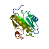
| ||||||||
|---|---|---|---|---|---|---|---|---|---|
| 1 |
| ||||||||
| Unit cell |
| ||||||||
| Atom site foot note | 1: CIS PROLINE - PRO 131 |
-Components
| #1: Protein | Mass: 16344.479 Da / Num. of mol.: 1 Source method: isolated from a genetically manipulated source Source: (gene. exp.) Enterobacteria phage T7 (virus) / Genus: T7-like viruses / Gene: T7 / Plasmid: T7 / Gene (production host): T7References: UniProt: P00806, N-acetylmuramoyl-L-alanine amidase |
|---|---|
| #2: Chemical | ChemComp-ZN /   Mass: 65.409 Da / Num. of mol.: 1 / Source method: obtained synthetically / Formula: Zn Mass: 65.409 Da / Num. of mol.: 1 / Source method: obtained synthetically / Formula: Zn |
| #3: Water | ChemComp-HOH / Water Mass: 18.015 Da / Num. of mol.: 26 / Source method: isolated from a natural source / Formula: H2O Mass: 18.015 Da / Num. of mol.: 26 / Source method: isolated from a natural source / Formula: H2O |
-Experimental details
-Experiment
| Experiment | Method: X-RAY DIFFRACTION |
|---|
- Sample preparation
| Crystal | Density Matthews: 2.44 Å3/Da / Density % sol: 49.68 % | ||||||||||||||||||||||||||||||||||||||||||||||||||||||||||||||||||||||||
|---|---|---|---|---|---|---|---|---|---|---|---|---|---|---|---|---|---|---|---|---|---|---|---|---|---|---|---|---|---|---|---|---|---|---|---|---|---|---|---|---|---|---|---|---|---|---|---|---|---|---|---|---|---|---|---|---|---|---|---|---|---|---|---|---|---|---|---|---|---|---|---|---|---|
| Crystal grow | *PLUS pH: 7 / Method: unknown | ||||||||||||||||||||||||||||||||||||||||||||||||||||||||||||||||||||||||
| Components of the solutions | *PLUS
|
-Data collection
| Radiation | Scattering type: x-ray |
|---|---|
| Radiation wavelength | Relative weight: 1 |
| Reflection | *PLUS Highest resolution: 2.2 Å / Num. obs: 7142 / Num. measured all: 42968 / Rmerge(I) obs: 0.0861 |
- Processing
| Software |
| ||||||||||||||||||||||||||||||||||||||||||||||||||||||||||||
|---|---|---|---|---|---|---|---|---|---|---|---|---|---|---|---|---|---|---|---|---|---|---|---|---|---|---|---|---|---|---|---|---|---|---|---|---|---|---|---|---|---|---|---|---|---|---|---|---|---|---|---|---|---|---|---|---|---|---|---|---|---|
| Refinement | Resolution: 2.2→10 Å / σ(F): 2 /
| ||||||||||||||||||||||||||||||||||||||||||||||||||||||||||||
| Refinement step | Cycle: LAST / Resolution: 2.2→10 Å
| ||||||||||||||||||||||||||||||||||||||||||||||||||||||||||||
| Refine LS restraints |
| ||||||||||||||||||||||||||||||||||||||||||||||||||||||||||||
| Software | *PLUS Name: X-PLOR / Classification: refinement | ||||||||||||||||||||||||||||||||||||||||||||||||||||||||||||
| Refinement | *PLUS Rfactor obs: 0.19 | ||||||||||||||||||||||||||||||||||||||||||||||||||||||||||||
| Solvent computation | *PLUS | ||||||||||||||||||||||||||||||||||||||||||||||||||||||||||||
| Displacement parameters | *PLUS | ||||||||||||||||||||||||||||||||||||||||||||||||||||||||||||
| Refine LS restraints | *PLUS Type: x_angle_d / Dev ideal: 3.3 |
