 Movie
Movie Controller

[English] 日本語
 Yorodumi
- PDB-1l7e: Crystal Structure of R. rubrum Transhydrogenase Domain I with Bou... -
+ Open data

- Basic information
| Entry | Database: PDB / ID: 1l7e | ||||||
|---|---|---|---|---|---|---|---|
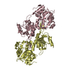
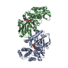
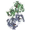
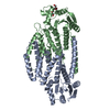
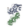
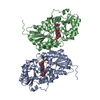
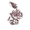
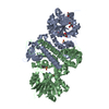
| Title | Crystal Structure of R. rubrum Transhydrogenase Domain I with Bound NADH | ||||||
 Components Components | nicotinamide nucleotide Transhydrogenase, subunit alpha 1 | ||||||
 Keywords Keywords | OXIDOREDUCTASE / Transhydrogenase domain I with NADH bound | ||||||
| Function / homology |  Function and homology informationNAD(P)+ transhydrogenase (B-specific) activity / NAD(P)+ transhydrogenase (AB-specific) activity / proton-translocating NAD(P)+ transhydrogenase / NADPH regeneration / NADH binding / NAD+ binding / NAD binding / protein dimerization activity Function and homology informationNAD(P)+ transhydrogenase (B-specific) activity / NAD(P)+ transhydrogenase (AB-specific) activity / proton-translocating NAD(P)+ transhydrogenase / NADPH regeneration / NADH binding / NAD+ binding / NAD binding / protein dimerization activitySimilarity search - Function | ||||||
| Biological species |  Rhodospirillum rubrum (bacteria) Rhodospirillum rubrum (bacteria) | ||||||
| Method | X-RAY DIFFRACTION / SYNCHROTRON / MOLECULAR REPLACEMENT / Resolution: 1.9 Å | ||||||
 Authors Authors | Prasad, G.S. / Wahlberg, M. / Sridhar, V. / Yamaguchi, M. / Hatefi, Y. / Stout, C.D. | ||||||
 Citation Citation | Journal: Biochemistry / Year: 2002 Title: Crystal Structures of Transhydrogenase Domain I with and without Bound NADH Authors: Prasad, G.S. / Wahlberg, M. / Sridhar, V. / Yamaguchi, M. / Hatefi, Y. / Stout, C.D. | ||||||
| History |
|
- Structure visualization
| Structure viewer | Molecule: MolmilJmol/JSmol |
|---|
- Downloads & links
-Download
| PDBx/mmCIF format | 1l7e.cif.gz | 289.4 KB | Display | PDBx/mmCIF format |
|---|---|---|---|---|
| PDB format | pdb1l7e.ent.gz | 228.5 KB | Display | PDB format |
| PDBx/mmJSON format | 1l7e.json.gz | Tree view | PDBx/mmJSON format | |
| Others |  Other downloads Other downloads |
-Validation report
| Arichive directory | https://data.pdbj.org/pub/pdb/validation_reports/l7/1l7eftp://data.pdbj.org/pub/pdb/validation_reports/l7/1l7e | HTTPS FTP |
|---|
-Related structure data
| Related structure data |  1l7dSC S: Starting model for refinement C: citing same article ( |
|---|---|
| Similar structure data |

-Links
 PDBj
PDBj

- Assembly
| Deposited unit | 
| ||||||||
|---|---|---|---|---|---|---|---|---|---|
| 1 | 
| ||||||||
| 2 | 
| ||||||||
| Unit cell |
|
-Components
| #1: Protein | / Transhydrogenase Domain I Mass: 40324.785 Da / Num. of mol.: 4 Source method: isolated from a genetically manipulated source Source: (gene. exp.) Rhodospirillum rubrum (bacteria) / Production host: Escherichia coli (E. coli)References: UniProt: Q60164, UniProt: Q2RSB2*PLUS, EC: 1.6.1.1 #2: Chemical | Nicotinamide adenine dinucleotide  Mass: 665.441 Da / Num. of mol.: 2 / Source method: obtained synthetically / Formula: C21H29N7O14P2 Mass: 665.441 Da / Num. of mol.: 2 / Source method: obtained synthetically / Formula: C21H29N7O14P2#3: Water | ChemComp-HOH / | Water Mass: 18.015 Da / Num. of mol.: 607 / Source method: isolated from a natural source / Formula: H2O Mass: 18.015 Da / Num. of mol.: 607 / Source method: isolated from a natural source / Formula: H2O |
|---|
-Experimental details
-Experiment
| Experiment | Method: X-RAY DIFFRACTION / Number of used crystals: 1 |
|---|
- Sample preparation
| Crystal | Density Matthews: 2.06 Å3/Da / Density % sol: 40.32 % | ||||||||||||||||||||||||||||||||||||||||||||||||||||||
|---|---|---|---|---|---|---|---|---|---|---|---|---|---|---|---|---|---|---|---|---|---|---|---|---|---|---|---|---|---|---|---|---|---|---|---|---|---|---|---|---|---|---|---|---|---|---|---|---|---|---|---|---|---|---|---|
| Crystal grow | Temperature: 277 K / Method: vapor diffusion / pH: 7.5 Details: mPEG 2K, Tris-HCl, Magnesium acetate, NADH, pH 7.5, VAPOR DIFFUSION, temperature 277.0K | ||||||||||||||||||||||||||||||||||||||||||||||||||||||
| Crystal grow | *PLUS Temperature: 4 ℃ / pH: 8 | ||||||||||||||||||||||||||||||||||||||||||||||||||||||
| Components of the solutions | *PLUS
|
-Data collection
| Diffraction | Mean temperature: 100 K |
|---|---|
| Diffraction source | Source: SYNCHROTRON / Site: SSRL  / Beamline: BL11-1 / Wavelength: 0.965 Å / Beamline: BL11-1 / Wavelength: 0.965 Å |
| Detector | Type: ADSC QUANTUM 4 / Detector: CCD / Date: Apr 21, 2001 |
| Radiation | Monochromator: Curved crystal (silicon 111) / Protocol: SINGLE WAVELENGTH / Monochromatic (M) / Laue (L): M / Scattering type: x-ray |
| Radiation wavelength | Wavelength: 0.965 Å / Relative weight: 1 |
| Reflection | Resolution: 1.9→50 Å / Num. all: 326796 / Num. obs: 102189 / % possible obs: 99.4 % / Observed criterion σ(F): 1.5 / Observed criterion σ(I): 0 / Redundancy: 3.2 % / Biso Wilson estimate: 30.9 Å2 / Rsym value: 0.119 / Net I/σ(I): 9.1 |
| Reflection shell | Resolution: 1.9→1.95 Å / Redundancy: 3 % / Mean I/σ(I) obs: 1.5 / Rsym value: 0.375 / % possible all: 99.4 |
| Reflection | *PLUS Num. measured all: 326796 / Rmerge(I) obs: 0.119 |
| Reflection shell | *PLUS Rmerge(I) obs: 0.375 |
- Processing
| Software |
| ||||||||||||||||||||
|---|---|---|---|---|---|---|---|---|---|---|---|---|---|---|---|---|---|---|---|---|---|
| Refinement | Method to determine structure: MOLECULAR REPLACEMENT Starting model: 1L7D Resolution: 1.9→50 Å / σ(F): 0 / Stereochemistry target values: Engh & Huber
| ||||||||||||||||||||
| Refinement step | Cycle: LAST / Resolution: 1.9→50 Å
| ||||||||||||||||||||
| Refine LS restraints |
| ||||||||||||||||||||
| Refinement | *PLUS | ||||||||||||||||||||
| Solvent computation | *PLUS | ||||||||||||||||||||
| Displacement parameters | *PLUS |
