 Movie
Movie Controller

[English] 日本語
 Yorodumi
- PDB-1f8g: THE X-RAY STRUCTURE OF NICOTINAMIDE NUCLEOTIDE TRANSHYDROGENASE F... -
+ Open data

- Basic information
| Entry | Database: PDB / ID: 1f8g | ||||||
|---|---|---|---|---|---|---|---|
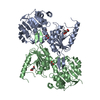
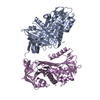
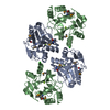
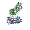
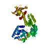
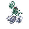
| Title | THE X-RAY STRUCTURE OF NICOTINAMIDE NUCLEOTIDE TRANSHYDROGENASE FROM RHODOSPIRILLUM RUBRUM COMPLEXED WITH NAD+ | ||||||
 Components Components | NICOTINAMIDE NUCLEOTIDE TRANSHYDROGENASE | ||||||
 Keywords Keywords | OXIDOREDUCTASE / Nucleotide fold / Proton pump Transhydrogenase / Rossmann fold | ||||||
| Function / homology |  Function and homology information Function and homology informationNAD(P)+ transhydrogenase (Si-specific) activity / proton-translocating NAD(P)+ transhydrogenase activity / proton-translocating NAD(P)+ transhydrogenase / NADH binding / NADPH regeneration / NAD+ binding / NAD binding / NADP binding / protein dimerization activity / plasma membrane Similarity search - Function | ||||||
| Biological species |  Rhodospirillum rubrum (bacteria) Rhodospirillum rubrum (bacteria) | ||||||
| Method |  X-RAY DIFFRACTION / SYNCHROTRON / MAD / Resolution: 2 Å X-RAY DIFFRACTION / SYNCHROTRON / MAD / Resolution: 2 Å | ||||||
 Authors Authors | Buckley, P.A. / Baz Jackson, J. / Schneider, T. / White, S.A. / Rice, D.W. / Baker, P.J. | ||||||
 Citation Citation | Journal: Structure Fold.Des. / Year: 2000 Title: Protein-protein recognition, hydride transfer and proton pumping in the transhydrogenase complex. Authors: Buckley, P.A. / Baz Jackson, J. / Schneider, T. / White, S.A. / Rice, D.W. / Baker, P.J. #1: Journal: To be PublishedTitle: The Crystallization of the dI Component of Transhydrogenase, A Proton-Translocating Membrane Protein Authors: Sedelnikova, S.E. / Burke, J. / Buckley, P.A. / Rice, D.W. / Jackson, J.B. / Cotton, N.P.J. / Grindley, R.L. / Baker, P.J. | ||||||
| History |
|
- Structure visualization
| Structure viewer | Molecule: MolmilJmol/JSmol |
|---|
- Downloads & links
-Download
| PDBx/mmCIF format | 1f8g.cif.gz | 323.8 KB | Display | PDBx/mmCIF format |
|---|---|---|---|---|
| PDB format | pdb1f8g.ent.gz | 261.3 KB | Display | PDB format |
| PDBx/mmJSON format | 1f8g.json.gz | Tree view | PDBx/mmJSON format | |
| Others |  Other downloads Other downloads |
-Validation report
| Arichive directory | https://data.pdbj.org/pub/pdb/validation_reports/f8/1f8gftp://data.pdbj.org/pub/pdb/validation_reports/f8/1f8g | HTTPS FTP |
|---|
-Related structure data
| Similar structure data |
|---|

-Links
 PDBj
PDBj- Assembly
| Deposited unit | 
| ||||||||
|---|---|---|---|---|---|---|---|---|---|
| 1 | 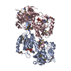
| ||||||||
| 2 | 
| ||||||||
| Unit cell |
| ||||||||
| Details | The biological assembly is a dimer. Two are present in asymmetric unit, chains A and C and chains B and D. |
-Components
| #1: Protein | Mass: 41028.203 Da / Num. of mol.: 4 Source method: isolated from a genetically manipulated source Source: (gene. exp.) Rhodospirillum rubrum (bacteria) / Plasmid: PCD1 / Production host: References: UniProt: Q60164, UniProt: Q2RSB2*PLUS, NAD(P)+ transhydrogenase (Si-specific) #2: Chemical | ChemComp-NAD /   Mass: 663.425 Da / Num. of mol.: 4 / Source method: obtained synthetically / Formula: C21H27N7O14P2 / Comment: NAD*YM Mass: 663.425 Da / Num. of mol.: 4 / Source method: obtained synthetically / Formula: C21H27N7O14P2 / Comment: NAD*YM#3: Water | ChemComp-HOH / |  Mass: 18.015 Da / Num. of mol.: 1459 / Source method: isolated from a natural source / Formula: H2O Mass: 18.015 Da / Num. of mol.: 1459 / Source method: isolated from a natural source / Formula: H2OHas protein modification | Y | |
|---|
-Experimental details
-Experiment
| Experiment | Method: X-RAY DIFFRACTION |
|---|
- Sample preparation
| Crystal | Density Matthews: 2.36 Å3/Da / Density % sol: 46.84 % | ||||||||||||||||||||||||||||||||||||||||
|---|---|---|---|---|---|---|---|---|---|---|---|---|---|---|---|---|---|---|---|---|---|---|---|---|---|---|---|---|---|---|---|---|---|---|---|---|---|---|---|---|---|
| Crystal grow | Temperature: 290 K / Method: vapor diffusion, hanging drop / pH: 7.5 Details: 10mM NAD (+), 25% methylethyl PEG 2000, 200mM sodium phosphate buffer, pH7.5, 30mM Ammonium sulphate. , VAPOR DIFFUSION, HANGING DROP, temperature 290K | ||||||||||||||||||||||||||||||||||||||||
| Crystal grow | *PLUS Details: Sedelnikova, S.E., (2000) Acta Crystallogr., D56, 1170. | ||||||||||||||||||||||||||||||||||||||||
| Components of the solutions | *PLUS
|
-Data collection
| Diffraction |
| ||||||||||||||||||||
|---|---|---|---|---|---|---|---|---|---|---|---|---|---|---|---|---|---|---|---|---|---|
| Diffraction source |
| ||||||||||||||||||||
| Detector |
| ||||||||||||||||||||
| Radiation | Protocol: SINGLE WAVELENGTH / Monochromatic (M) / Laue (L): M / Scattering type: x-ray | ||||||||||||||||||||
| Radiation wavelength |
| ||||||||||||||||||||
| Reflection | Resolution: 2→20 Å / Num. all: 99140 / Num. obs: 99140 / % possible obs: 94.7 % / Redundancy: 3.22 % / Biso Wilson estimate: 23.3 Å2 / Rmerge(I) obs: 0.043 / Net I/σ(I): 14.1 | ||||||||||||||||||||
| Reflection shell | Resolution: 2→2.05 Å / Redundancy: 2.6 % / Rmerge(I) obs: 0.271 / Num. unique all: 5966 / % possible all: 89.4 | ||||||||||||||||||||
| Reflection | *PLUS | ||||||||||||||||||||
| Reflection shell | *PLUS Mean I/σ(I) obs: 2.5 |

- Processing
| Software |
| ||||||||||||||||||||
|---|---|---|---|---|---|---|---|---|---|---|---|---|---|---|---|---|---|---|---|---|---|
| Refinement | Method to determine structure: MAD / Resolution: 2→20 Å / Cross valid method: THROUGHOUT / σ(F): 0 / σ(I): 0 / Stereochemistry target values: Engh and Huber
| ||||||||||||||||||||
| Refinement step | Cycle: LAST / Resolution: 2→20 Å
| ||||||||||||||||||||
| Refine LS restraints |
| ||||||||||||||||||||
| Software | *PLUS Name: REFMAC / Classification: refinement | ||||||||||||||||||||
| Refinement | *PLUS Highest resolution: 2 Å / σ(F): 0 / % reflection Rfree: 5 % / Rfactor obs: 0.21 / Rfactor Rfree: 0.26 / Rfactor Rwork: 0.21 | ||||||||||||||||||||
| Solvent computation | *PLUS | ||||||||||||||||||||
| Displacement parameters | *PLUS |
