 Movie
Movie Controller


+ Open data

- Basic information
| Entry | Database: PDB / ID: 1kv3 | ||||||
|---|---|---|---|---|---|---|---|
| Title | HUMAN TISSUE TRANSGLUTAMINASE IN GDP BOUND FORM | ||||||
 Components Components | Protein-glutamine gamma-glutamyltransferase | ||||||
 Keywords Keywords | TRANSFERASE / tissue transglutaminase / GTP binding protein / crystallography | ||||||
| Function / homology |  Function and homology information Function and homology informationprotein deamination / histone serotonyltransferase activity / histone dopaminyltransferase activity / peptide noradrenalinyltransferase activity / peptide histaminyltransferase activity / cellular response to serotonin / regulation of apoptotic cell clearance / protein-glutamine glutaminase activity / protein-glutamine glutaminase / protein-glutamine gamma-glutamyltransferase ...protein deamination / histone serotonyltransferase activity / histone dopaminyltransferase activity / peptide noradrenalinyltransferase activity / peptide histaminyltransferase activity / cellular response to serotonin / regulation of apoptotic cell clearance / protein-glutamine glutaminase activity / protein-glutamine glutaminase / protein-glutamine gamma-glutamyltransferase / positive regulation of mitochondrial calcium ion concentration / salivary gland cavitation / protein-glutamine gamma-glutamyltransferase activity / negative regulation of endoplasmic reticulum calcium ion concentration / dopamine secretion / peptide cross-linking / branching involved in salivary gland morphogenesis / cellular response to dopamine / positive regulation of small GTPase mediated signal transduction / cellular response to cocaine / apoptotic cell clearance / Hydrolases; Acting on peptide bonds (peptidases) / positive regulation of neurogenesis / positive regulation of sprouting angiogenesis / positive regulation of GTPase activity / Transferases; Acyltransferases; Transferring groups other than aminoacyl groups / positive regulation of cell adhesion / protein homooligomerization / bone development / extracellular matrix / : / peptidase activity / nucleosome / gene expression / regulation of apoptotic process / phospholipase C-activating G protein-coupled receptor signaling pathway / positive regulation of apoptotic process / focal adhesion / calcium ion binding / GTP binding / chromatin / perinuclear region of cytoplasm / endoplasmic reticulum / mitochondrion / proteolysis / extracellular exosome / nucleus / plasma membrane / cytosol Similarity search - Function | ||||||
| Biological species |  Homo sapiens (human) Homo sapiens (human) | ||||||
| Method |  X-RAY DIFFRACTION / SYNCHROTRON / MOLECULAR REPLACEMENT / Resolution: 2.8 Å X-RAY DIFFRACTION / SYNCHROTRON / MOLECULAR REPLACEMENT / Resolution: 2.8 Å | ||||||
 Authors Authors | Liu, S. / Cerione, R.A. / Clardy, J. | ||||||
 Citation Citation | Journal: Proc.Natl.Acad.Sci.USA / Year: 2002 Title: Structural basis for the guanine nucleotide-binding activity of tissue transglutaminase and its regulation of transamidation activity. Authors: Liu, S. / Cerione, R.A. / Clardy, J. | ||||||
| History |
| ||||||
| Remark 999 | SEQUENCE According to Swissprot entry P21980, residues 51, 186, 224, 533 and 655 are in conflict. ...SEQUENCE According to Swissprot entry P21980, residues 51, 186, 224, 533 and 655 are in conflict. Residue 51 can be either GLU or GLN, Residue 186 GLU or GLN, residue 224 either VAL or GLY, residue 533 either ASN or THR, and residue 655 can be either LEU or VAL. |
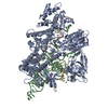
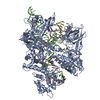
- Structure visualization
| Structure viewer | Molecule: MolmilJmol/JSmol |
|---|
- Downloads & links
-Download
| PDBx/mmCIF format | 1kv3.cif.gz | 765.5 KB | Display | PDBx/mmCIF format |
|---|---|---|---|---|
| PDB format | pdb1kv3.ent.gz | 635.1 KB | Display | PDB format |
| PDBx/mmJSON format | 1kv3.json.gz | Tree view | PDBx/mmJSON format | |
| Others |  Other downloads Other downloads |
-Validation report
| Arichive directory | https://data.pdbj.org/pub/pdb/validation_reports/kv/1kv3ftp://data.pdbj.org/pub/pdb/validation_reports/kv/1kv3 | HTTPS FTP |
|---|
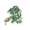
-Related structure data
| Related structure data |  1gguS S: Starting model for refinement |
|---|---|
| Similar structure data |


-Links
 PDBj
PDBj
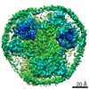
- Assembly
| Deposited unit | 
| ||||||||
|---|---|---|---|---|---|---|---|---|---|
| 1 | 
| ||||||||
| 2 | 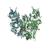
| ||||||||
| 3 | 
| ||||||||
| Unit cell |
|
-Components
| #1: Protein | Mass: 77341.602 Da / Num. of mol.: 6 Source method: isolated from a genetically manipulated source Source: (gene. exp.) Homo sapiens (human) / Gene: TGM2 / Plasmid: PET28a / Species (production host): Escherichia coli / Production host:  References: UniProt: P21980, protein-glutamine gamma-glutamyltransferase #2: Chemical | ChemComp-GDP /   Type: RNA linking / Mass: 443.201 Da / Num. of mol.: 6 / Source method: obtained synthetically / Formula: C10H15N5O11P2 / Comment: GDP, energy-carrying molecule*YM Type: RNA linking / Mass: 443.201 Da / Num. of mol.: 6 / Source method: obtained synthetically / Formula: C10H15N5O11P2 / Comment: GDP, energy-carrying molecule*YM#3: Water | ChemComp-HOH / |  Mass: 18.015 Da / Num. of mol.: 428 / Source method: isolated from a natural source / Formula: H2O Mass: 18.015 Da / Num. of mol.: 428 / Source method: isolated from a natural source / Formula: H2O |
|---|
-Experimental details
-Experiment
| Experiment | Method: X-RAY DIFFRACTION / Number of used crystals: 1 |
|---|
- Sample preparation
| Crystal | Density Matthews: 2.87 Å3/Da / Density % sol: 57.19 % | |||||||||||||||||||||||||||||||||||||||||||||||||||||||||||||||||||||||||||||
|---|---|---|---|---|---|---|---|---|---|---|---|---|---|---|---|---|---|---|---|---|---|---|---|---|---|---|---|---|---|---|---|---|---|---|---|---|---|---|---|---|---|---|---|---|---|---|---|---|---|---|---|---|---|---|---|---|---|---|---|---|---|---|---|---|---|---|---|---|---|---|---|---|---|---|---|---|---|---|
| Crystal grow | Temperature: 277 K / Method: vapor diffusion, hanging drop / pH: 6.6 Details: 50 mM MES, pH 6.6, 200 mM NaCl, 50 mM MgCl2, 6-8% PEG3350 and 5 mM DTT, VAPOR DIFFUSION, HANGING DROP, temperature 4K | |||||||||||||||||||||||||||||||||||||||||||||||||||||||||||||||||||||||||||||
| Crystal grow | *PLUS pH: 7 / Method: vapor diffusion, sitting drop | |||||||||||||||||||||||||||||||||||||||||||||||||||||||||||||||||||||||||||||
| Components of the solutions | *PLUS
|
-Data collection
| Diffraction | Mean temperature: 100 K |
|---|---|
| Diffraction source | Source: SYNCHROTRON / Site: APS  / Beamline: 14-BM-D / Wavelength: 1 Å / Beamline: 14-BM-D / Wavelength: 1 Å |
| Detector | Type: ADSC QUANTUM 4 / Detector: CCD / Date: Nov 29, 2000 / Details: mirros |
| Radiation | Monochromator: mirror / Protocol: SINGLE WAVELENGTH / Monochromatic (M) / Laue (L): M / Scattering type: x-ray |
| Radiation wavelength | Wavelength: 1 Å / Relative weight: 1 |
| Reflection | Resolution: 2.8→52 Å / Num. all: 131998 / Num. obs: 124870 / % possible obs: 94.6 % / Observed criterion σ(F): 0 / Observed criterion σ(I): 0 / Redundancy: 4.4 % / Biso Wilson estimate: 64.6 Å2 / Rmerge(I) obs: 0.085 / Rsym value: 0.085 |
| Reflection shell | Resolution: 2.8→2.98 Å / Redundancy: 4 % / Rmerge(I) obs: 0.47 / Num. unique all: 18358 / Rsym value: 0.47 / % possible all: 88.8 |
| Reflection | *PLUS Lowest resolution: 51.8 Å / Num. measured all: 1141553 |
| Reflection shell | *PLUS % possible obs: 88.8 % / Rmerge(I) obs: 0.47 |
- Processing
| Software |
| ||||||||||||||||||||||||||||||||||||
|---|---|---|---|---|---|---|---|---|---|---|---|---|---|---|---|---|---|---|---|---|---|---|---|---|---|---|---|---|---|---|---|---|---|---|---|---|---|
| Refinement | Method to determine structure: MOLECULAR REPLACEMENT Starting model: factor XIIIa, PDB entry 1GGU Resolution: 2.8→51.79 Å / Rfactor Rfree error: 0.003 / Data cutoff high absF: 3021702.44 / Data cutoff low absF: 0 / Isotropic thermal model: RESTRAINED / Cross valid method: THROUGHOUT / σ(F): 0 / σ(I): 0 / Stereochemistry target values: Engh & Huber Details: Non-crystallographic symmetry restrain used on 6 copies of molecules in the asymmetric unit
| ||||||||||||||||||||||||||||||||||||
| Solvent computation | Solvent model: FLAT MODEL / Bsol: 14.1343 Å2 / ksol: 0.271762 e/Å3 | ||||||||||||||||||||||||||||||||||||
| Displacement parameters | Biso mean: 57.2 Å2
| ||||||||||||||||||||||||||||||||||||
| Refine analyze |
| ||||||||||||||||||||||||||||||||||||
| Refinement step | Cycle: LAST / Resolution: 2.8→51.79 Å
| ||||||||||||||||||||||||||||||||||||
| Refine LS restraints |
| ||||||||||||||||||||||||||||||||||||
| LS refinement shell | Resolution: 2.8→2.98 Å / Rfactor Rfree error: 0.013 / Total num. of bins used: 6
| ||||||||||||||||||||||||||||||||||||
| Xplor file |
| ||||||||||||||||||||||||||||||||||||
| Refinement | *PLUS | ||||||||||||||||||||||||||||||||||||
| Solvent computation | *PLUS | ||||||||||||||||||||||||||||||||||||
| Displacement parameters | *PLUS | ||||||||||||||||||||||||||||||||||||
| Refine LS restraints | *PLUS
| ||||||||||||||||||||||||||||||||||||
| LS refinement shell | *PLUS Rfactor Rwork: 0.36 |
