 Movie
Movie Controller


+ Open data

- Basic information
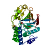
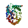
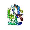
| Entry | Database: PDB / ID: 1kr0 | |||||||||
|---|---|---|---|---|---|---|---|---|---|---|
| Title | Hevamine Mutant D125A/Y183F in Complex with Tetra-NAG | |||||||||
 Components Components | Hevamine A | |||||||||
 Keywords Keywords | HYDROLASE / chitinase/lysozyme | |||||||||
| Function / homology |  Function and homology information Function and homology informationchitinase activity / endochitinase activity / chitinase / vacuole / chitin catabolic process / polysaccharide catabolic process / lysozyme / lysozyme activity / extracellular region Similarity search - Function | |||||||||
| Biological species |   Hevea brasiliensis (rubber tree) Hevea brasiliensis (rubber tree) | |||||||||
| Method |  X-RAY DIFFRACTION / FOURIER SYNTHESIS / Resolution: 1.92 Å X-RAY DIFFRACTION / FOURIER SYNTHESIS / Resolution: 1.92 Å | |||||||||
 Authors Authors | Rozeboom, H.J. / Dijkstra, B.W. | |||||||||
 Citation Citation | Journal: Eur.J.Biochem. / Year: 2002 Title: Expression and Characterization of Active Site Mutants of Hevamine, a Chitinase from the Rubber Tree Hevea brasiliensis. Authors: Bokma, E. / Rozeboom, H.J. / Sibbald, M. / Dijkstra, B.W. / Beintema, J.J. #1: Journal: J.Mol.Biol. / Year: 1996Title: The 1.8 A Resolution Structure of Hevamine, a Plant Chitinase/Lysozyme, and Analysis of the Conserved Sequence and Structure Motifs of Glycosyl Hydrolase Family 18. Authors: Terwisscha van Scheltinga, A.C. / Hennig, M. / Dijkstra, B.W. #2: Journal: Biochemistry / Year: 1995Title: Stereochemistry of Chitin Hydrolysis by a Plant Chitinase/Lysozyme and X-ray Structure of a Complex with Allosamidin: Evidence for Substrate Assisted Catalysis. Authors: Terwisscha van Scheltinga, A.C. / Armand, S. / Kalk, K.H. / Isogai, A. / Henrissat, B. / Dijkstra, B.W. #3: Journal: Structure / Year: 1994Title: Crystal Structures of Hevamine, a Plant Defence Protein with Chitinase and Lysozyme Activity, and its Complex with an Inhibitor. Authors: Terwisscha van Scheltinga, A.C. / Kalk, K.H. / Beintema, J.J. / Dijkstra, B.W. #4: Journal: J.Mol.Biol. / Year: 1990Title: Crystallization of Hevamine, an Enzyme with Lysozyme/Chitinase Activity from Hevea brasiliensis Latex. Authors: Rozeboom, H.J. / Budiani, A. / Beintema, J.J. / Dijkstra, B.W. | |||||||||
| History |
|
- Structure visualization
| Structure viewer | Molecule: MolmilJmol/JSmol |
|---|
- Downloads & links
-Download
| PDBx/mmCIF format | 1kr0.cif.gz | 69.4 KB | Display | PDBx/mmCIF format |
|---|---|---|---|---|
| PDB format | pdb1kr0.ent.gz | 50.6 KB | Display | PDB format |
| PDBx/mmJSON format | 1kr0.json.gz | Tree view | PDBx/mmJSON format | |
| Others |  Other downloads Other downloads |
-Validation report
| Arichive directory | https://data.pdbj.org/pub/pdb/validation_reports/kr/1kr0ftp://data.pdbj.org/pub/pdb/validation_reports/kr/1kr0 | HTTPS FTP |
|---|
-Related structure data
| Related structure data |  1kqyC  1kqzC  1kr1C  2hvmS S: Starting model for refinement C: citing same article ( |
|---|---|
| Similar structure data |


-Links
 PDBj
PDBj- Assembly
| Deposited unit | 
| ||||||||
|---|---|---|---|---|---|---|---|---|---|
| 1 |
| ||||||||
| Unit cell |
|
-Components
| #1: Protein | Mass: 29513.174 Da / Num. of mol.: 1 / Mutation: D125A/Y183F Source method: isolated from a genetically manipulated source Source: (gene. exp.) Hevea brasiliensis (rubber tree) / Plasmid: pGELAF+ / Production host:  References: UniProt: p23472, UniProt: P23472*PLUS, chitinase, lysozyme |
|---|---|
| #2: Polysaccharide | 2-acetamido-2-deoxy-beta-D-glucopyranose-(1-4)-2-acetamido-2-deoxy-beta-D-glucopyranose-(1-4)-2- ...2-acetamido-2-deoxy-beta-D-glucopyranose-(1-4)-2-acetamido-2-deoxy-beta-D-glucopyranose-(1-4)-2-acetamido-2-deoxy-beta-D-glucopyranose-(1-4)-2-acetamido-2-deoxy-beta-D-glucopyranose Source method: isolated from a genetically manipulated source |
| #3: Chemical | ChemComp-SO4 /   Mass: 96.063 Da / Num. of mol.: 1 / Source method: obtained synthetically / Formula: SO4 Mass: 96.063 Da / Num. of mol.: 1 / Source method: obtained synthetically / Formula: SO4 |
| #4: Water | ChemComp-HOH /  Mass: 18.015 Da / Num. of mol.: 142 / Source method: isolated from a natural source / Formula: H2O Mass: 18.015 Da / Num. of mol.: 142 / Source method: isolated from a natural source / Formula: H2O |
| Has protein modification | Y |
-Experimental details
-Experiment
| Experiment | Method: X-RAY DIFFRACTION / Number of used crystals: 1 |
|---|
- Sample preparation
| Crystal | Density Matthews: 2.2 Å3/Da / Density % sol: 44 % |
|---|---|
| Crystal grow | Temperature: 293 K / Method: vapor diffusion, hanging drop / pH: 7 Details: Ammonium sulfate, pH 7.0, VAPOR DIFFUSION, HANGING DROP, temperature 293K |
| Crystal grow | *PLUS Method: unknown |
| Components of the solutions | *PLUS Conc.: 1.1-1.4 M / Common name: ammonium sulfate / Details: or 10-30%(w/v) PEG3350, pH7.0 |
-Data collection
| Diffraction | Mean temperature: 293 K |
|---|---|
| Diffraction source | Source: ROTATING ANODE / Type: ENRAF-NONIUS / Wavelength: 1.5418 Å |
| Detector | Type: MACSCIENCE / Detector: IMAGE PLATE / Date: Feb 14, 2000 |
| Radiation | Protocol: SINGLE WAVELENGTH / Monochromatic (M) / Laue (L): M / Scattering type: x-ray |
| Radiation wavelength | Wavelength: 1.5418 Å / Relative weight: 1 |
| Reflection | Resolution: 1.92→50 Å / Num. all: 19371 / Num. obs: 19371 / % possible obs: 99.8 % / Observed criterion σ(I): -3 / Redundancy: 9.7 % / Biso Wilson estimate: 18.6 Å2 / Rmerge(I) obs: 0.087 / Net I/σ(I): 14.8 |
| Reflection shell | Resolution: 1.92→1.95 Å / Rmerge(I) obs: 0.324 / Mean I/σ(I) obs: 4.5 / Num. unique all: 927 / % possible all: 97.4 |
| Reflection | *PLUS Lowest resolution: 44 Å / Num. obs: 19419 / Num. measured all: 187850 |
| Reflection shell | *PLUS % possible obs: 97.4 % |
- Processing
| Software |
| ||||||||||||||||||||||||||||||||||||||||||||||||||||||||||||||||||||||||||||||||
|---|---|---|---|---|---|---|---|---|---|---|---|---|---|---|---|---|---|---|---|---|---|---|---|---|---|---|---|---|---|---|---|---|---|---|---|---|---|---|---|---|---|---|---|---|---|---|---|---|---|---|---|---|---|---|---|---|---|---|---|---|---|---|---|---|---|---|---|---|---|---|---|---|---|---|---|---|---|---|---|---|---|
| Refinement | Method to determine structure: FOURIER SYNTHESIS Starting model: 2HVM Resolution: 1.92→43.95 Å / Rfactor Rfree error: 0.006 / Data cutoff high absF: 1274359.78 / Data cutoff low absF: 0 / Isotropic thermal model: RESTRAINED / Cross valid method: THROUGHOUT / σ(F): 0 / Stereochemistry target values: Engh & Huber
| ||||||||||||||||||||||||||||||||||||||||||||||||||||||||||||||||||||||||||||||||
| Solvent computation | Solvent model: FLAT MODEL / Bsol: 49.2003 Å2 / ksol: 0.352757 e/Å3 | ||||||||||||||||||||||||||||||||||||||||||||||||||||||||||||||||||||||||||||||||
| Displacement parameters | Biso mean: 22.4 Å2
| ||||||||||||||||||||||||||||||||||||||||||||||||||||||||||||||||||||||||||||||||
| Refine analyze |
| ||||||||||||||||||||||||||||||||||||||||||||||||||||||||||||||||||||||||||||||||
| Refinement step | Cycle: LAST / Resolution: 1.92→43.95 Å
| ||||||||||||||||||||||||||||||||||||||||||||||||||||||||||||||||||||||||||||||||
| Refine LS restraints |
| ||||||||||||||||||||||||||||||||||||||||||||||||||||||||||||||||||||||||||||||||
| LS refinement shell | Resolution: 1.92→1.99 Å / Rfactor Rfree error: 0.026 / Total num. of bins used: 10
| ||||||||||||||||||||||||||||||||||||||||||||||||||||||||||||||||||||||||||||||||
| Xplor file |
| ||||||||||||||||||||||||||||||||||||||||||||||||||||||||||||||||||||||||||||||||
| Software | *PLUS Name: CNS / Version: 1 / Classification: refinement | ||||||||||||||||||||||||||||||||||||||||||||||||||||||||||||||||||||||||||||||||
| Refinement | *PLUS σ(F): 0 / % reflection Rfree: 5.2 % / Rfactor obs: 0.167 / Rfactor Rfree: 0.202 | ||||||||||||||||||||||||||||||||||||||||||||||||||||||||||||||||||||||||||||||||
| Solvent computation | *PLUS | ||||||||||||||||||||||||||||||||||||||||||||||||||||||||||||||||||||||||||||||||
| Displacement parameters | *PLUS Biso mean: 22.4 Å2 | ||||||||||||||||||||||||||||||||||||||||||||||||||||||||||||||||||||||||||||||||
| Refine LS restraints | *PLUS
| ||||||||||||||||||||||||||||||||||||||||||||||||||||||||||||||||||||||||||||||||
| LS refinement shell | *PLUS Rfactor Rfree: 0.247 / % reflection Rfree: 5.2 % / Rfactor Rwork: 0.212 |
