 Movie
Movie Controller

[English] 日本語
 Yorodumi
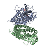
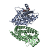
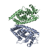
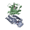
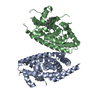
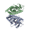
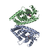
- PDB-1knu: LIGAND BINDING DOMAIN OF THE HUMAN PEROXISOME PROLIFERATOR ACTIVA... -
+ Open data

- Basic information
| Entry | Database: PDB / ID: 1knu | ||||||
|---|---|---|---|---|---|---|---|
| Title | LIGAND BINDING DOMAIN OF THE HUMAN PEROXISOME PROLIFERATOR ACTIVATED RECEPTOR GAMMA IN COMPLEX WITH A SYNTHETIC AGONIST | ||||||
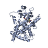
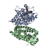
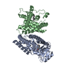
 Components Components | PEROXISOME PROLIFERATOR ACTIVATED RECEPTOR GAMMA | ||||||
 Keywords Keywords | DNA BINDING PROTEIN / PPAR / NUCLEAR RECEPTOR / TRANSCRIPTION / GENE REGULATION / AGONIST COMPLEX | ||||||
| Function / homology |  Function and homology information Function and homology informationprostaglandin receptor activity / beige fat cell differentiation / negative regulation of receptor signaling pathway via STAT / MECP2 regulates transcription factors / negative regulation of vascular endothelial cell proliferation / negative regulation of extracellular matrix assembly / negative regulation of connective tissue replacement involved in inflammatory response wound healing / positive regulation of cholesterol transport / negative regulation of cellular response to transforming growth factor beta stimulus / arachidonate binding ...prostaglandin receptor activity / beige fat cell differentiation / negative regulation of receptor signaling pathway via STAT / MECP2 regulates transcription factors / negative regulation of vascular endothelial cell proliferation / negative regulation of extracellular matrix assembly / negative regulation of connective tissue replacement involved in inflammatory response wound healing / positive regulation of cholesterol transport / negative regulation of cellular response to transforming growth factor beta stimulus / arachidonate binding / positive regulation of adiponectin secretion / DNA binding domain binding / positive regulation of vascular associated smooth muscle cell apoptotic process / negative regulation of cardiac muscle hypertrophy in response to stress / positive regulation of fatty acid metabolic process / WW domain binding / STAT family protein binding / positive regulation of lipid metabolic process / response to lipid / negative regulation of type II interferon-mediated signaling pathway / LBD domain binding / negative regulation of cholesterol storage / positive regulation of lipoprotein transport / negative regulation of SMAD protein signal transduction / lipid homeostasis / E-box binding / alpha-actinin binding / R-SMAD binding / negative regulation of vascular associated smooth muscle cell proliferation / negative regulation of blood vessel endothelial cell migration / white fat cell differentiation / negative regulation of macrophage derived foam cell differentiation / negative regulation of lipid storage / positive regulation of cholesterol efflux / monocyte differentiation / negative regulation of BMP signaling pathway / cell fate commitment / cellular response to low-density lipoprotein particle stimulus / negative regulation of mitochondrial fission / negative regulation of osteoblast differentiation / long-chain fatty acid transport / positive regulation of fat cell differentiation / BMP signaling pathway / nuclear retinoid X receptor binding / fat cell differentiation / Transcriptional regulation of brown and beige adipocyte differentiation by EBF2 / retinoic acid receptor signaling pathway / negative regulation of MAPK cascade / intracellular receptor signaling pathway / cell maturation / positive regulation of adipose tissue development / hormone-mediated signaling pathway / peroxisome proliferator activated receptor signaling pathway / epithelial cell differentiation / regulation of cellular response to insulin stimulus / peptide binding / response to nutrient / brown fat cell differentiation / negative regulation of miRNA transcription / negative regulation of angiogenesis / placenta development / Regulation of PTEN gene transcription / transcription coregulator binding / positive regulation of apoptotic signaling pathway / SUMOylation of intracellular receptors / negative regulation of smooth muscle cell proliferation / negative regulation of transforming growth factor beta receptor signaling pathway / PPARA activates gene expression / fatty acid metabolic process / regulation of circadian rhythm / Nuclear Receptor transcription pathway / Transcriptional regulation of white adipocyte differentiation / mRNA transcription by RNA polymerase II / positive regulation of miRNA transcription / negative regulation of inflammatory response / DNA-binding transcription repressor activity, RNA polymerase II-specific / regulation of blood pressure / RNA polymerase II transcription regulator complex / nuclear receptor activity / cellular response to insulin stimulus / rhythmic process / glucose homeostasis / MLL4 and MLL3 complexes regulate expression of PPARG target genes in adipogenesis and hepatic steatosis / DNA-binding transcription activator activity, RNA polymerase II-specific / double-stranded DNA binding / cellular response to hypoxia / sequence-specific DNA binding / DNA-binding transcription factor binding / nucleic acid binding / DNA-binding transcription factor activity, RNA polymerase II-specific / cell differentiation / receptor complex / transcription cis-regulatory region binding / RNA polymerase II cis-regulatory region sequence-specific DNA binding / DNA-binding transcription factor activity / negative regulation of gene expression / innate immune response / negative regulation of DNA-templated transcription / chromatin binding / positive regulation of gene expression Similarity search - Function | ||||||
| Biological species |  Homo sapiens (human) Homo sapiens (human) | ||||||
| Method |  X-RAY DIFFRACTION / SYNCHROTRON / FOURIER SYNTHESIS / Resolution: 2.5 Å X-RAY DIFFRACTION / SYNCHROTRON / FOURIER SYNTHESIS / Resolution: 2.5 Å | ||||||
 Authors Authors | Svensson, L.A. / Mortensen, S.B. / Fleckner, J. / Woeldike, H.F. | ||||||
 Citation Citation | Journal: J.MED.CHEM. / Year: 2002 Title: Novel tricyclic-alpha-alkyloxyphenylpropionic acids: dual PPARalpha/gamma agonists with hypolipidemic and antidiabetic activity Authors: Sauerberg, P. / Pettersson, I. / Jeppesen, L. / Bury, P.S. / Mogensen, J.P. / Wassermann, K. / Brand, C.L. / Sturis, J. / Woldike, H.F. / Fleckner, J. / Andersen, A.-S.T. / Mortensen, S.B. / ...Authors: Sauerberg, P. / Pettersson, I. / Jeppesen, L. / Bury, P.S. / Mogensen, J.P. / Wassermann, K. / Brand, C.L. / Sturis, J. / Woldike, H.F. / Fleckner, J. / Andersen, A.-S.T. / Mortensen, S.B. / Svensson, L.A. / Rasmussen, H.B. / Lehmann, S.V. / Polivka, Z. / Sindelar, K. / Panajotova, V. / Ynddal, L. / Wulff, E.M. | ||||||
| History |
|
- Structure visualization
| Structure viewer | Molecule: MolmilJmol/JSmol |
|---|
- Downloads & links
-Download
| PDBx/mmCIF format | 1knu.cif.gz | 115.9 KB | Display | PDBx/mmCIF format |
|---|---|---|---|---|
| PDB format | pdb1knu.ent.gz | 91 KB | Display | PDB format |
| PDBx/mmJSON format | 1knu.json.gz | Tree view | PDBx/mmJSON format | |
| Others |  Other downloads Other downloads |
-Validation report
| Arichive directory | https://data.pdbj.org/pub/pdb/validation_reports/kn/1knuftp://data.pdbj.org/pub/pdb/validation_reports/kn/1knu | HTTPS FTP |
|---|
-Related structure data
| Related structure data | 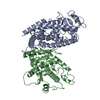 1prgS S: Starting model for refinement |
|---|---|
| Similar structure data |

-Links
 PDBj
PDBj- Assembly
| Deposited unit | 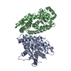
| ||||||||
|---|---|---|---|---|---|---|---|---|---|
| 1 |
| ||||||||
| Unit cell |
|
-Components
| #1: Protein | Mass: 31321.393 Da / Num. of mol.: 2 / Fragment: LIGAND BINDING DOMAIN Source method: isolated from a genetically manipulated source Source: (gene. exp.) Homo sapiens (human) / Tissue: ADIPOSE TISSUE / Gene: PPARG, NR1C3 / Plasmid: pGEX-3X-hPPARgLBD / Species (production host): Escherichia coli / Production host:  #2: Chemical | ChemComp-YPA / ( | 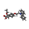  Mass: 403.470 Da / Num. of mol.: 1 / Source method: obtained synthetically / Formula: C25H25NO4 Mass: 403.470 Da / Num. of mol.: 1 / Source method: obtained synthetically / Formula: C25H25NO4#3: Water | ChemComp-HOH / |  Mass: 18.015 Da / Num. of mol.: 32 / Source method: isolated from a natural source / Formula: H2O Mass: 18.015 Da / Num. of mol.: 32 / Source method: isolated from a natural source / Formula: H2O |
|---|
-Experimental details
-Experiment
| Experiment | Method: X-RAY DIFFRACTION / Number of used crystals: 1 |
|---|
- Sample preparation
| Crystal | Density Matthews: 2.73 Å3/Da / Density % sol: 54.87 % | ||||||||||||||||||||||||
|---|---|---|---|---|---|---|---|---|---|---|---|---|---|---|---|---|---|---|---|---|---|---|---|---|---|
| Crystal grow | Temperature: 294 K / Method: vapor diffusion, sitting drop / pH: 8 Details: CITRIC ACID, TRIS-HCL, pH 8.0, VAPOR DIFFUSION, SITTING DROP, temperature 294.0K | ||||||||||||||||||||||||
| Crystal grow | *PLUS Method: unknown | ||||||||||||||||||||||||
| Components of the solutions | *PLUS
|
-Data collection
| Diffraction | Mean temperature: 100 K |
|---|---|
| Diffraction source | Source: SYNCHROTRON / Site: MAX II  / Beamline: I711 / Wavelength: 1.0292 Å / Beamline: I711 / Wavelength: 1.0292 Å |
| Detector | Type: MARRESEARCH / Detector: IMAGE PLATE / Date: Sep 14, 2000 |
| Radiation | Monochromator: SAGITALLY FOCUSED Si(111) / Protocol: SINGLE WAVELENGTH / Monochromatic (M) / Laue (L): M / Scattering type: x-ray |
| Radiation wavelength | Wavelength: 1.0292 Å / Relative weight: 1 |
| Reflection | Resolution: 2.5→40 Å / Num. all: 22943 / Num. obs: 22943 / % possible obs: 97.3 % / Observed criterion σ(F): 0 / Observed criterion σ(I): -3 / Redundancy: 3.2 % / Biso Wilson estimate: 49.8 Å2 / Rmerge(I) obs: 0.076 / Net I/σ(I): 13.05 |
| Reflection shell | Resolution: 2.5→2.59 Å / Redundancy: 2.7 % / Rmerge(I) obs: 0.343 / Mean I/σ(I) obs: 3.49 / Num. unique all: 2293 / % possible all: 98 |
| Reflection shell | *PLUS % possible obs: 98.2 % / Mean I/σ(I) obs: 3.5 |
- Processing
| Software |
| ||||||||||||||||||||||||||||||||||||||||||||||||||||||||||||||||||||||||||||||||
|---|---|---|---|---|---|---|---|---|---|---|---|---|---|---|---|---|---|---|---|---|---|---|---|---|---|---|---|---|---|---|---|---|---|---|---|---|---|---|---|---|---|---|---|---|---|---|---|---|---|---|---|---|---|---|---|---|---|---|---|---|---|---|---|---|---|---|---|---|---|---|---|---|---|---|---|---|---|---|---|---|---|
| Refinement | Method to determine structure: FOURIER SYNTHESIS Starting model: PDB ENTRY 1PRG Resolution: 2.5→39.89 Å / Rfactor Rfree error: 0.008 / Data cutoff high absF: 4042063.32 / Data cutoff low absF: 0 / Isotropic thermal model: RESTRAINED / Cross valid method: THROUGHOUT / σ(F): 0 / Stereochemistry target values: Engh & Huber
| ||||||||||||||||||||||||||||||||||||||||||||||||||||||||||||||||||||||||||||||||
| Displacement parameters | Biso mean: 57.2 Å2
| ||||||||||||||||||||||||||||||||||||||||||||||||||||||||||||||||||||||||||||||||
| Refine analyze |
| ||||||||||||||||||||||||||||||||||||||||||||||||||||||||||||||||||||||||||||||||
| Refinement step | Cycle: LAST / Resolution: 2.5→39.89 Å
| ||||||||||||||||||||||||||||||||||||||||||||||||||||||||||||||||||||||||||||||||
| Refine LS restraints |
| ||||||||||||||||||||||||||||||||||||||||||||||||||||||||||||||||||||||||||||||||
| LS refinement shell | Resolution: 2.5→2.59 Å / Rfactor Rfree error: 0.035 / Total num. of bins used: 10
| ||||||||||||||||||||||||||||||||||||||||||||||||||||||||||||||||||||||||||||||||
| Xplor file |
| ||||||||||||||||||||||||||||||||||||||||||||||||||||||||||||||||||||||||||||||||
| Refinement | *PLUS Lowest resolution: 40 Å | ||||||||||||||||||||||||||||||||||||||||||||||||||||||||||||||||||||||||||||||||
| Solvent computation | *PLUS | ||||||||||||||||||||||||||||||||||||||||||||||||||||||||||||||||||||||||||||||||
| Displacement parameters | *PLUS | ||||||||||||||||||||||||||||||||||||||||||||||||||||||||||||||||||||||||||||||||
| Refine LS restraints | *PLUS Type: c_bond_d / Dev ideal: 0.008 |
