 Movie
Movie Controller

[English] 日本語
 Yorodumi
- PDB-1k0e: THE CRYSTAL STRUCTURE OF AMINODEOXYCHORISMATE SYNTHASE FROM FORMA... -
+ Open data

- Basic information
| Entry | Database: PDB / ID: 1k0e | ||||||
|---|---|---|---|---|---|---|---|
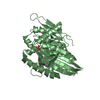
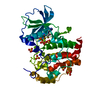
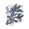
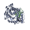
| Title | THE CRYSTAL STRUCTURE OF AMINODEOXYCHORISMATE SYNTHASE FROM FORMATE GROWN CRYSTALS | ||||||
 Components Components | p-aminobenzoate synthase component I | ||||||
 Keywords Keywords | LYASE / AMINODEOXYCHORISMATE SYNTHASE / CHORISMATE / GLUTAMINE / TRYPTOPHAN / PABA SYNTHASE / P-AMINOBENZOATE SYNTHASE | ||||||
| Function / homology |  Function and homology information Function and homology informationaminodeoxychorismate synthase complex / aminodeoxychorismate synthase / tryptophan binding / 4-amino-4-deoxychorismate synthase activity / 4-aminobenzoate biosynthetic process / L-tryptophan biosynthetic process / folic acid biosynthetic process / tetrahydrofolate biosynthetic process / protein heterodimerization activity / magnesium ion binding Similarity search - Function | ||||||
| Biological species |  | ||||||
| Method |  X-RAY DIFFRACTION / SYNCHROTRON / MAD / Resolution: 2 Å X-RAY DIFFRACTION / SYNCHROTRON / MAD / Resolution: 2 Å | ||||||
 Authors Authors | Parsons, J.F. / Jensen, P.Y. / Pachikara, A.S. / Howard, A.J. / Eisenstein, E. / Ladner, J.E. | ||||||
 Citation Citation | Journal: Biochemistry / Year: 2002 Title: Structure of Escherichia coli aminodeoxychorismate synthase: architectural conservation and diversity in chorismate-utilizing enzymes. Authors: Parsons, J.F. / Jensen, P.Y. / Pachikara, A.S. / Howard, A.J. / Eisenstein, E. / Ladner, J.E. | ||||||
| History |
|
- Structure visualization
| Structure viewer | Molecule: MolmilJmol/JSmol |
|---|
- Downloads & links
-Download
| PDBx/mmCIF format | 1k0e.cif.gz | 190.7 KB | Display | PDBx/mmCIF format |
|---|---|---|---|---|
| PDB format | pdb1k0e.ent.gz | 146.7 KB | Display | PDB format |
| PDBx/mmJSON format | 1k0e.json.gz | Tree view | PDBx/mmJSON format | |
| Others |  Other downloads Other downloads |
-Validation report
| Arichive directory | https://data.pdbj.org/pub/pdb/validation_reports/k0/1k0eftp://data.pdbj.org/pub/pdb/validation_reports/k0/1k0e | HTTPS FTP |
|---|
-Related structure data


-Links
 PDBj
PDBj
- Assembly
| Deposited unit | 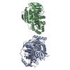
| ||||||||
|---|---|---|---|---|---|---|---|---|---|
| 1 | 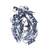
| ||||||||
| 2 | 
| ||||||||
| Unit cell |
|
-Components
| #1: Protein | Mass: 51022.316 Da / Num. of mol.: 2 Source method: isolated from a genetically manipulated source Source: (gene. exp.) References: UniProt: P05041, Lyases; Carbon-carbon lyases; Oxo-acid-lyases #2: Chemical |   Type: L-peptide linking / Mass: 204.225 Da / Num. of mol.: 2 / Source method: obtained synthetically / Formula: C11H12N2O2 Type: L-peptide linking / Mass: 204.225 Da / Num. of mol.: 2 / Source method: obtained synthetically / Formula: C11H12N2O2#3: Chemical | ChemComp-FMT / |   Mass: 46.025 Da / Num. of mol.: 1 / Source method: obtained synthetically / Formula: CH2O2 Mass: 46.025 Da / Num. of mol.: 1 / Source method: obtained synthetically / Formula: CH2O2#4: Water | ChemComp-HOH / |  Mass: 18.015 Da / Num. of mol.: 318 / Source method: isolated from a natural source / Formula: H2O Mass: 18.015 Da / Num. of mol.: 318 / Source method: isolated from a natural source / Formula: H2O |
|---|
-Experimental details
-Experiment
| Experiment | Method: X-RAY DIFFRACTION / Number of used crystals: 1 |
|---|
- Sample preparation
| Crystal | Density Matthews: 2.1 Å3/Da / Density % sol: 41.4 % | ||||||||||||||||||||||||||||||||||||||||||||||||||||||||
|---|---|---|---|---|---|---|---|---|---|---|---|---|---|---|---|---|---|---|---|---|---|---|---|---|---|---|---|---|---|---|---|---|---|---|---|---|---|---|---|---|---|---|---|---|---|---|---|---|---|---|---|---|---|---|---|---|---|
| Crystal grow | Temperature: 298 K / Method: vapor diffusion, hanging drop / pH: 4.6 Details: 0.1M sodium acetate buffer, 2.0M sodium formate, pH 4.6, VAPOR DIFFUSION, HANGING DROP, temperature 298K (PROTEIN SOLUTION: 50mM MOPS pH 7.5, 50mM KCL, 5mM MG CL2, 2 mM DTT, 40.2 MG/ML ...Details: 0.1M sodium acetate buffer, 2.0M sodium formate, pH 4.6, VAPOR DIFFUSION, HANGING DROP, temperature 298K (PROTEIN SOLUTION: 50mM MOPS pH 7.5, 50mM KCL, 5mM MG CL2, 2 mM DTT, 40.2 MG/ML PROTEIN. WELL SOLUTION: 0.1 M NA ACETATE pH 4.6, 2.0 M NA FORMATE) | ||||||||||||||||||||||||||||||||||||||||||||||||||||||||
| Components of the solutions |
| ||||||||||||||||||||||||||||||||||||||||||||||||||||||||
| Crystal grow | *PLUS pH: 7.4 | ||||||||||||||||||||||||||||||||||||||||||||||||||||||||
| Components of the solutions | *PLUS
|
-Data collection
| Diffraction | Mean temperature: 115 K | |||||||||
|---|---|---|---|---|---|---|---|---|---|---|
| Diffraction source | Source: SYNCHROTRON / Site: APS  / Beamline: 17-BM / Wavelength: 1 / Wavelength: 1 Å / Beamline: 17-BM / Wavelength: 1 / Wavelength: 1 Å | |||||||||
| Detector | Type: MARRESEARCH / Detector: CCD / Date: Nov 17, 2000 | |||||||||
| Radiation | Protocol: SINGLE WAVELENGTH / Monochromatic (M) / Laue (L): M / Scattering type: x-ray | |||||||||
| Radiation wavelength |
| |||||||||
| Reflection | Resolution: 2→20 Å / Num. all: 269253 / Num. obs: 55584 / % possible obs: 91.2 % / Redundancy: 5 % / Rmerge(I) obs: 0.1 / Net I/σ(I): 11.5 | |||||||||
| Reflection shell | Resolution: 2→2.08 Å / Redundancy: 3 % / Rmerge(I) obs: 0.208 / Mean I/σ(I) obs: 4.8 / % possible all: 90.4 | |||||||||
| Reflection | *PLUS Highest resolution: 2 Å / Lowest resolution: 20 Å / Redundancy: 5 % / Num. measured all: 269253 / Rmerge(I) obs: 0.1 | |||||||||
| Reflection shell | *PLUS % possible obs: 90.4 % / Redundancy: 3 % |
- Processing
| Software |
| |||||||||||||||||||||||||||||||||
|---|---|---|---|---|---|---|---|---|---|---|---|---|---|---|---|---|---|---|---|---|---|---|---|---|---|---|---|---|---|---|---|---|---|---|
| Refinement | Method to determine structure: MAD / Resolution: 2→10 Å / Num. parameters: 28479 / Num. restraintsaints: 28146 / Cross valid method: FREE R / σ(F): 0 / Stereochemistry target values: ENGH AND HUBER Details: ANISOTROPIC SCALING APPLIED BY THE METHOD OF PARKIN, MOEZZI & HOPE, J.APPL.CRYST.28(1995)53-56
| |||||||||||||||||||||||||||||||||
| Solvent computation | Solvent model: MOEWS & KRETSINGER, J.MOL.BIOL.91(1973)201-228 | |||||||||||||||||||||||||||||||||
| Refine analyze | Num. disordered residues: 0 / Occupancy sum hydrogen: 0 / Occupancy sum non hydrogen: 7116 | |||||||||||||||||||||||||||||||||
| Refinement step | Cycle: LAST / Resolution: 2→10 Å
| |||||||||||||||||||||||||||||||||
| Refine LS restraints |
| |||||||||||||||||||||||||||||||||
| Software | *PLUS Name: SHELXL-97 / Classification: refinement | |||||||||||||||||||||||||||||||||
| Refinement | *PLUS Lowest resolution: 10 Å / σ(F): 0 / % reflection Rfree: 5 % / Rfactor Rwork: 0.205 / Rfactor Rfree: 0.315 | |||||||||||||||||||||||||||||||||
| Solvent computation | *PLUS | |||||||||||||||||||||||||||||||||
| Displacement parameters | *PLUS | |||||||||||||||||||||||||||||||||
| Refine LS restraints | *PLUS Type: s_plane_restr / Dev ideal: 0.018 |
