 Movie
Movie Controller


+ Open data

- Basic information
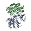
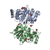
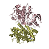
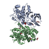
| Entry | Database: PDB / ID: 1jow | ||||||
|---|---|---|---|---|---|---|---|
| Title | Crystal structure of a complex of human CDK6 and a viral cyclin | ||||||
 Components Components |
| ||||||
 Keywords Keywords | CELL CYCLE/TRANSFERASE / CDK-cyclin complex / cyclin fold / CELL CYCLE-TRANSFERASE COMPLEX | ||||||
| Function / homology |  Function and homology information Function and homology informationcyclin D2-CDK6 complex / cyclin D3-CDK6 complex / cyclin D1-CDK6 complex / cell dedifferentiation / Evasion of Oncogene Induced Senescence Due to Defective p16INK4A binding to CDK4 and CDK6 / Evasion of Oxidative Stress Induced Senescence Due to Defective p16INK4A binding to CDK4 and CDK6 / Drug-mediated inhibition of CDK4/CDK6 activity / FBXO family protein binding / lateral ventricle development / symbiont-mediated perturbation of host cell cycle progression ...cyclin D2-CDK6 complex / cyclin D3-CDK6 complex / cyclin D1-CDK6 complex / cell dedifferentiation / Evasion of Oncogene Induced Senescence Due to Defective p16INK4A binding to CDK4 and CDK6 / Evasion of Oxidative Stress Induced Senescence Due to Defective p16INK4A binding to CDK4 and CDK6 / Drug-mediated inhibition of CDK4/CDK6 activity / FBXO family protein binding / lateral ventricle development / symbiont-mediated perturbation of host cell cycle progression / negative regulation of myeloid cell differentiation / type B pancreatic cell development / negative regulation of monocyte differentiation / astrocyte development / dentate gyrus development / regulation of cell motility / gliogenesis / Regulation of RUNX1 Expression and Activity / regulation of hematopoietic stem cell differentiation / positive regulation of cell-matrix adhesion / generation of neurons / Defective binding of RB1 mutants to E2F1,(E2F2, E2F3) / negative regulation of cell cycle / negative regulation of cellular senescence / negative regulation of cell differentiation / hematopoietic stem cell differentiation / cyclin-dependent kinase / negative regulation of osteoblast differentiation / cyclin-dependent protein serine/threonine kinase activity / cyclin-dependent protein kinase holoenzyme complex / regulation of G2/M transition of mitotic cell cycle / Notch signaling pathway / ruffle / cyclin binding / regulation of erythrocyte differentiation / G1/S transition of mitotic cell cycle / Oncogene Induced Senescence / response to virus / positive regulation of fibroblast proliferation / negative regulation of epithelial cell proliferation / Cyclin D associated events in G1 / T cell differentiation in thymus / regulation of gene expression / Senescence-Associated Secretory Phenotype (SASP) / Oxidative Stress Induced Senescence / regulation of cell cycle / negative regulation of cell population proliferation / cell division / protein serine kinase activity / DNA damage response / positive regulation of gene expression / centrosome / negative regulation of transcription by RNA polymerase II / signal transduction / nucleoplasm / ATP binding / nucleus / cytoplasm / cytosol Similarity search - Function | ||||||
| Biological species |  Saimiriine herpesvirus 2 (Herpesvirus saimiri) Saimiriine herpesvirus 2 (Herpesvirus saimiri) Homo sapiens (human) Homo sapiens (human) | ||||||
| Method |  X-RAY DIFFRACTION / SYNCHROTRON / MOLECULAR REPLACEMENT / Resolution: 3.1 Å X-RAY DIFFRACTION / SYNCHROTRON / MOLECULAR REPLACEMENT / Resolution: 3.1 Å | ||||||
 Authors Authors | Schulze-Gahmen, U. / Kim, S.H. | ||||||
 Citation Citation | Journal: Nat.Struct.Biol. / Year: 2002 Title: Structural basis for CDK6 activation by a virus-encoded cyclin. Authors: Schulze-Gahmen, U. / Kim, S.H. #1: Journal: To be PublishedTitle: Crystallization of a Complex between Human CDK6 and a Virus-encoded Cyclin is Critically Dependent on the Addition of Small Charged Organic Molecules Authors: Schulze-Gahmen, U. / Kim, S.H. | ||||||
| History |
|
- Structure visualization
| Structure viewer | Molecule: MolmilJmol/JSmol |
|---|
- Downloads & links
-Download
| PDBx/mmCIF format | 1jow.cif.gz | 102.3 KB | Display | PDBx/mmCIF format |
|---|---|---|---|---|
| PDB format | pdb1jow.ent.gz | 78.9 KB | Display | PDB format |
| PDBx/mmJSON format | 1jow.json.gz | Tree view | PDBx/mmJSON format | |
| Others |  Other downloads Other downloads |
-Validation report
| Arichive directory | https://data.pdbj.org/pub/pdb/validation_reports/jo/1jowftp://data.pdbj.org/pub/pdb/validation_reports/jo/1jow | HTTPS FTP |
|---|
-Related structure data
| Related structure data | |
|---|---|
| Similar structure data |


-Links
 PDBj
PDBj


- Assembly
| Deposited unit | 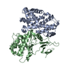
| ||||||||
|---|---|---|---|---|---|---|---|---|---|
| 1 |
| ||||||||
| Unit cell |
| ||||||||
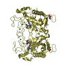
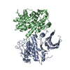
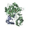
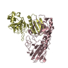
| Details | The biological assembly is the heterodimer in the asymmetric unit |
-Components
| #1: Protein | Mass: 28665.416 Da / Num. of mol.: 1 Source method: isolated from a genetically manipulated source Source: (gene. exp.) Saimiriine herpesvirus 2 (Herpesvirus saimiri)Genus: Rhadinovirus / Plasmid: pFastBac HTa donor plasmid / Cell line (production host): Sf9 / Production host:   Spodoptera frugiperda (fall armyworm) / References: UniProt: Q01043 Spodoptera frugiperda (fall armyworm) / References: UniProt: Q01043 |
|---|---|
| #2: Protein | Mass: 35066.332 Da / Num. of mol.: 1 / Fragment: residues 1-308 Source method: isolated from a genetically manipulated source Source: (gene. exp.) Homo sapiens (human) / Gene: CDK6 / Plasmid: pFastBac1 donor plasmid / Cell line (production host): Sf9 / Production host: Spodoptera frugiperda (fall armyworm)References: UniProt: Q00534, Transferases; Transferring phosphorus-containing groups; Phosphotransferases with an alcohol group as acceptor |
-Experimental details
-Experiment
| Experiment | Method: X-RAY DIFFRACTION / Number of used crystals: 1 |
|---|
- Sample preparation
| Crystal | Density Matthews: 2.5 Å3/Da / Density % sol: 50.78 % | |||||||||||||||||||||||||||||||||||||||||||||||||||||||||||||||
|---|---|---|---|---|---|---|---|---|---|---|---|---|---|---|---|---|---|---|---|---|---|---|---|---|---|---|---|---|---|---|---|---|---|---|---|---|---|---|---|---|---|---|---|---|---|---|---|---|---|---|---|---|---|---|---|---|---|---|---|---|---|---|---|---|
| Crystal grow | Temperature: 295 K / Method: vapor diffusion, hanging drop / pH: 8 Details: Tris/HCL, sodium chloride, PEG 3350, calcium acetate, pH 8.0, VAPOR DIFFUSION, HANGING DROP, temperature 295.0K | |||||||||||||||||||||||||||||||||||||||||||||||||||||||||||||||
| Crystal grow | *PLUS Temperature: 295 KDetails: Schulze-Gahmen, U., (2001) Acta Crystallogr, D57, 1287. | |||||||||||||||||||||||||||||||||||||||||||||||||||||||||||||||
| Components of the solutions | *PLUS
|
-Data collection
| Diffraction | Mean temperature: 100 K |
|---|---|
| Diffraction source | Source: SYNCHROTRON / Site: ALS  / Beamline: 5.0.2 / Wavelength: 0.99999 Å / Beamline: 5.0.2 / Wavelength: 0.99999 Å |
| Detector | Type: ADSC QUANTUM 4 / Detector: CCD / Date: Dec 17, 2000 |
| Radiation | Monochromator: double crystal / Protocol: SINGLE WAVELENGTH / Monochromatic (M) / Laue (L): M / Scattering type: x-ray |
| Radiation wavelength | Wavelength: 0.99999 Å / Relative weight: 1 |
| Reflection | Resolution: 3.1→30 Å / Num. all: 12815 / Num. obs: 12783 / % possible obs: 0.98 % / Observed criterion σ(F): 1 / Observed criterion σ(I): 1 / Redundancy: 7.8 % / Rsym value: 0.057 / Net I/σ(I): 29 |
| Reflection shell | Resolution: 3.1→3.19 Å / Mean I/σ(I) obs: 4 / Rsym value: 0.29 / % possible all: 0.82 |
| Reflection | *PLUS Num. obs: 12678 / % possible obs: 97.9 % / Num. measured all: 92567 / Rmerge(I) obs: 0.058 |
| Reflection shell | *PLUS % possible obs: 81.8 % / Rmerge(I) obs: 0.29 |
- Processing
| Software |
| |||||||||||||||||||||||||
|---|---|---|---|---|---|---|---|---|---|---|---|---|---|---|---|---|---|---|---|---|---|---|---|---|---|---|
| Refinement | Method to determine structure: MOLECULAR REPLACEMENT / Resolution: 3.1→19.93 Å / Rfactor Rfree error: 0.01 / Data cutoff high absF: 2293632.74 / Data cutoff low absF: 0 / Isotropic thermal model: GROUP / Cross valid method: THROUGHOUT / σ(F): 0 / Stereochemistry target values: engh & Huber
| |||||||||||||||||||||||||
| Solvent computation | Solvent model: FLAT MODEL / Bsol: 29.9899 Å2 / ksol: 0.272956 e/Å3 | |||||||||||||||||||||||||
| Displacement parameters | Biso mean: 73.3 Å2
| |||||||||||||||||||||||||
| Refine analyze |
| |||||||||||||||||||||||||
| Refinement step | Cycle: LAST / Resolution: 3.1→19.93 Å
| |||||||||||||||||||||||||
| Refine LS restraints |
| |||||||||||||||||||||||||
| LS refinement shell | Resolution: 3.1→3.29 Å / Rfactor Rfree error: 0.038 / Total num. of bins used: 6
| |||||||||||||||||||||||||
| Xplor file | Serial no: 1 / Param file: PROTEIN_REP.PARAM / Topol file: PROTEIN.TOP | |||||||||||||||||||||||||
| Software | *PLUS Name: CNS / Version: 1 / Classification: refinement | |||||||||||||||||||||||||
| Refinement | *PLUS Lowest resolution: 20 Å / σ(F): 0 / % reflection Rfree: 7.5 % / Rfactor obs: 0.266 | |||||||||||||||||||||||||
| Solvent computation | *PLUS | |||||||||||||||||||||||||
| Displacement parameters | *PLUS Biso mean: 73.3 Å2 | |||||||||||||||||||||||||
| Refine LS restraints | *PLUS
| |||||||||||||||||||||||||
| LS refinement shell | *PLUS Rfactor Rfree: 0.451 / % reflection Rfree: 7.8 % / Rfactor Rwork: 0.385 |
